Are you busy during this broadcast? See Broadcast #1 for an alternative time.
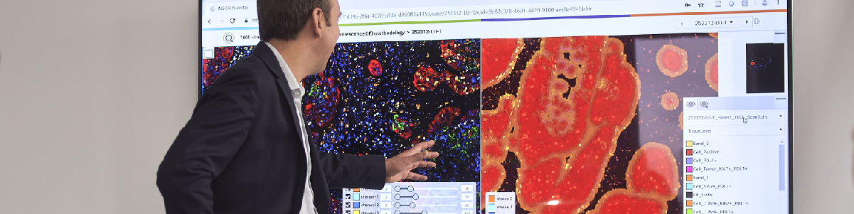
PD-L1 expression measured by immunohistochemistry helps identify non-small cell lung cancer (NSCLC) patients that may respond to anti-PD-1/PD-L1 immunotherapies. In this webinar, our featured speaker will present a novel deep learning solution for the automated scoring of PD-L1+ tumor cells (TC) in whole slide images of resected NSCLC.
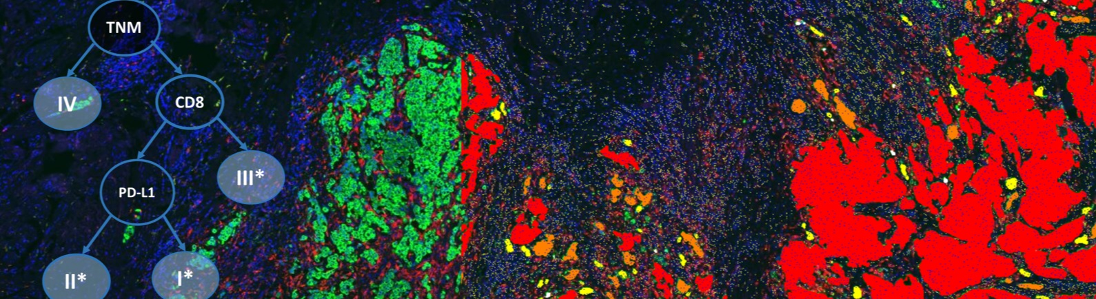
A convolutional neural network (CNN) was used for the fine-grained classification of tissue regions into three classes: (1) regions of membrane-positive epithelial tumor cells [TC(+)], (2) regions of membrane-negative epithelial tumor cells [TC(-)], and (3) other regions that could wrongly influence scoring, i.e. macrophages, positive and negative lymphocytes, stroma and/or necrosis. The TC score was calculated as the ratio of the area of the classified TC(+) region to the sum of the areas of the classified TC(-) and TC(+) regions.
Two sets of ~225k training and ~30k testing patches (128×128 pixels) were created from manual partial annotations from (N=22) train and (N=12) test slides (Ventana-SP263). Training a fully convolutional network yielded maximum accuracy of 0.89 on test patches. The trained network was applied on (N=433) unseen confirmation slides and the TC score calculated for each slide based on the classified TC(+) and TC(-) regions. A non-linear gamma mapping to the manual TC scores by a trained pathologist was then estimated to maximize Overall Percent Agreement (OPA) at ≥25% criterion using two-fold cross-validation.
Evaluation against pathologist scoring on the confirmation slides yielded higher Overall (OPA), Negative (NPA) and Positive (PPA) Percent Agreement values at ≥25% criterion, higher Lin’s correlation and lower mean absolute error than a baseline approach relying on a heuristic detection of individual epithelium cells [ESMO-2017-103P]. Scoring by a second pathologist was available on a subset (N=170) of the confirmation slides. Average and standard deviation results on this subset confirm the above observations and suggest that the approach is getting close to inter-pathologist variability.
Join this webinar to learn how using deep learning to identify PD-L1 positive and negative tumor cell regions enables the automated scoring of PD-L1 TCs at the ≥25% expression level in resected NSCLC. These findings should be confirmed with additional tumor sets.
Speaker

Nicolas Brieu, PhD, Senior Research Scientist, Definiens
Nicolas has more than 10 years’ experience in the field of medical image analysis. His main area of research is in machine and deep learning for computer vision and data mining. He joined Definiens in 2011 and has since been working on the research and development of novel algorithms to automate the analysis of digital pathology images and the identification of immune-related prognostic factors.
Who Should Attend?
This webinar will be ideal for medical, pharmaceutical, biotech and diagnostics executives, directors and vice presidents of therapeutic areas and Chief Scientific, Medical and Executive Officers. It will also be informative for lab and study directors, and medical directors.
What You Will Learn
Join this webinar to learn how using deep learning to identify PD-L1 positive and negative tumor cell regions enables the automated scoring of PD-L1 TCs at the ≥25% expression level in resected NSCLC.
Xtalks Partner

Definiens
We improve patient lives by unlocking the tissue phenome.
In oncology, therapeutic strategies have shifted from a direct assault on cancer cells to recruiting the immune system for that purpose. Our mission is to accelerate breakthroughs for this approach by helping scientists leverage Tissue Phenomics to deepen understanding of disease biology and immune system mechanisms, to bring multi-omics data into a cancer-relevant context, and to facilitate the translation of new insights into novel therapies and treatment strategies. Our vision is to create unique patient profiles for an individualized standard of care, where patients experience fewer side effects and live longer.
Definiens’ Tissue Phenomics approach was awarded the 2013 Frost and Sullivan Company of the Year Award for Global Tissue Diagnostics and Pathology Imaging. For more information, please visit: www.definiens.com.
Related Webinars
You Must Login To Register for this Free Webinar
Already have an account? LOGIN HERE. If you don’t have an account you need to create a free account.
Create Account