Non-alcoholic fatty liver disease (NAFLD) is the most common diffuse liver disease, with a worldwide prevalence of 20-46 percent. NAFLD can be subdivided into simple steatosis and nonalcoholic steatohepatitis (NASH).
Patients with non-alcoholic fatty liver disease (NAFLD) are at risk of steatohepatitis and progressive liver fibrosis culminating in cirrhosis, typically over a period of decades. Early diagnosis and risk stratification are essential for effective management.
Although liver biopsy is currently the gold standard for diagnosing progressive NASH, it has many drawbacks, such as sampling error, cost and risk of complications. Furthermore, it is not realistic to perform liver biopsies on all NAFLD patients.
The limitations of liver biopsy have driven a search for non-invasive NAFLD screening and risk stratification methods. Since advanced fibrosis has been proven to be prognostic of poor outcomes in NAFLD, multiple surrogate fibrosis markers have been studied, including imaging methods.
The two most prevalent imaging techniques to examine NAFLD and NASH patients are:
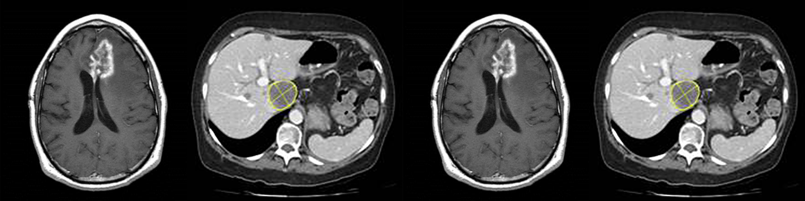
- Magnetic resonance elastography (MRE) — a technique that builds upon magnetic resonance imaging (MRI) to determine the stiffness of the liver, which can be used to determine the degree of fibrosis
- Proton density fat fraction (PDFF) — an MRI technique that can accurately determine the amount of fat in the liver (steatosis)
In this free webinar, the featured speaker will discuss these imaging methods, how they may be used for NAFLD/NASH screening, diagnosis & risk stratification, and other imaging techniques that may provide value for NAFLD/NASH patients.
Speaker

Jonathan Riek, PhD, VP, Musculoskeletal & Metabolic Imaging, BioTel Research
Jonathan Riek has a PhD in Electrical Engineering from the University of Rochester, and his doctoral thesis was on motion and artifact reduction in MRI. He worked on the development of a 3D CT scanner as a postdoctoral researcher, then spent seven years in the research labs at the Eastman Kodak Company. While at Kodak, Jonathan worked in the areas of motion estimation, frame rate conversion and video compression. He also served as the liaison between the image science division and the corporate software group. He joined VirtualScopics in 2000 to return to medical imaging and apply his skills interfacing between image science and software. In 2004, he led the development of the system that is still being used to run clinical trials and analyze medical images. In his current role, Jonathan has scientific responsibility for all of BioTel Research’s musculoskeletal and metabolic imaging studies, including arthritis, muscle diseases, diabetes and liver diseases such as NASH and NAFLD. He holds four patents and has published many articles on various topics regarding MRI.
Who Should Attend?
This webinar will benefit medical and non-medical professionals in the biopharmaceutical industry, especially those supporting NASH/NAFLD drug development with roles in:
- Clinical Research
- Clinical Development
- Medical Affairs
- Clinical Operations
- Project Management
- Regulatory Affairs
What You Will Learn
Participants will learn about:
- An overview of NASH/NAFLD
- Benefits and drawbacks of liver biopsy for liver disease diagnosis
- Non-invasive NAFLD screening and risk stratification
- Benefits and applications of two imaging technologies
- Magnetic Resonance Elastography (MRE)
- Proton Density Fat Fraction (PDFF)
Xtalks Partner
BioTel Research
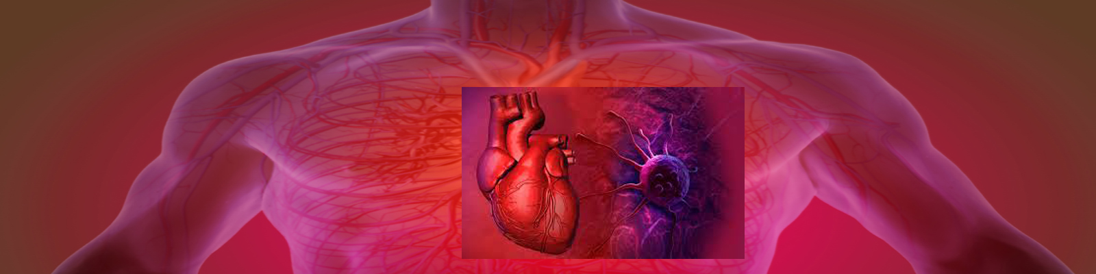
As an industry leader in testing services for clinical trials, BioTel Research combines the expert medical imaging of VirtualScopics and the cardiac core lab leadership of Cardiocore. BioTel Research offers global operational support for cardiovascular monitoring in all therapeutic areas, and advanced imaging services in oncology, cardiovascular, metabolic, musculoskeletal, neurologic and medical device studies. Their accessible and experienced research team comprises key opinion leaders, board-certified cardiologists and radiologists, sub-specialty scientists, and highly trained technicians — who acquire, evaluate, and report high-quality data through an efficient, cloud-based infrastructure. At BioTel Research, their job is to support your clinical trial with the most accessible and experienced team and the most advanced technologies available… through personal service on a global scale.
Related Webinars


You Must Login To Register for this Free Webinar
Already have an account? LOGIN HERE. If you don’t have an account you need to create a free account.
Create Account