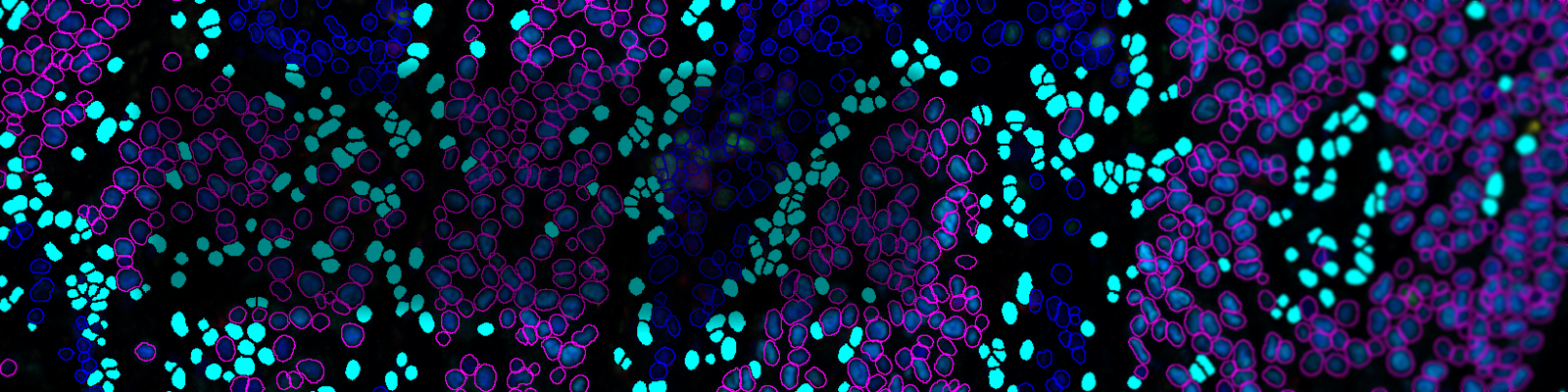
The immune response is spatially and temporally regulated. The density and location of immune cells in the tumor microenvironment (TME) have important diagnostic and prognostic values. Single-cell-based multi-omic technologies have exponentially increased our understanding of the numerous cellular and molecular networks regulating tumor initiation and progression. However, these techniques do not provide information about the spatial organization of cells or cell-cell interactions.
Affordable, accessible, and easy-to-execute multiplexing techniques that allow spatial resolution of immune cells in tissue sections are needed to complement single cell-based high-throughput technologies. Visiopharm® has developed a strategy that integrates serial imaging, sequential labeling, and image alignment to generate virtual multiparameter slides of whole tissue sections.
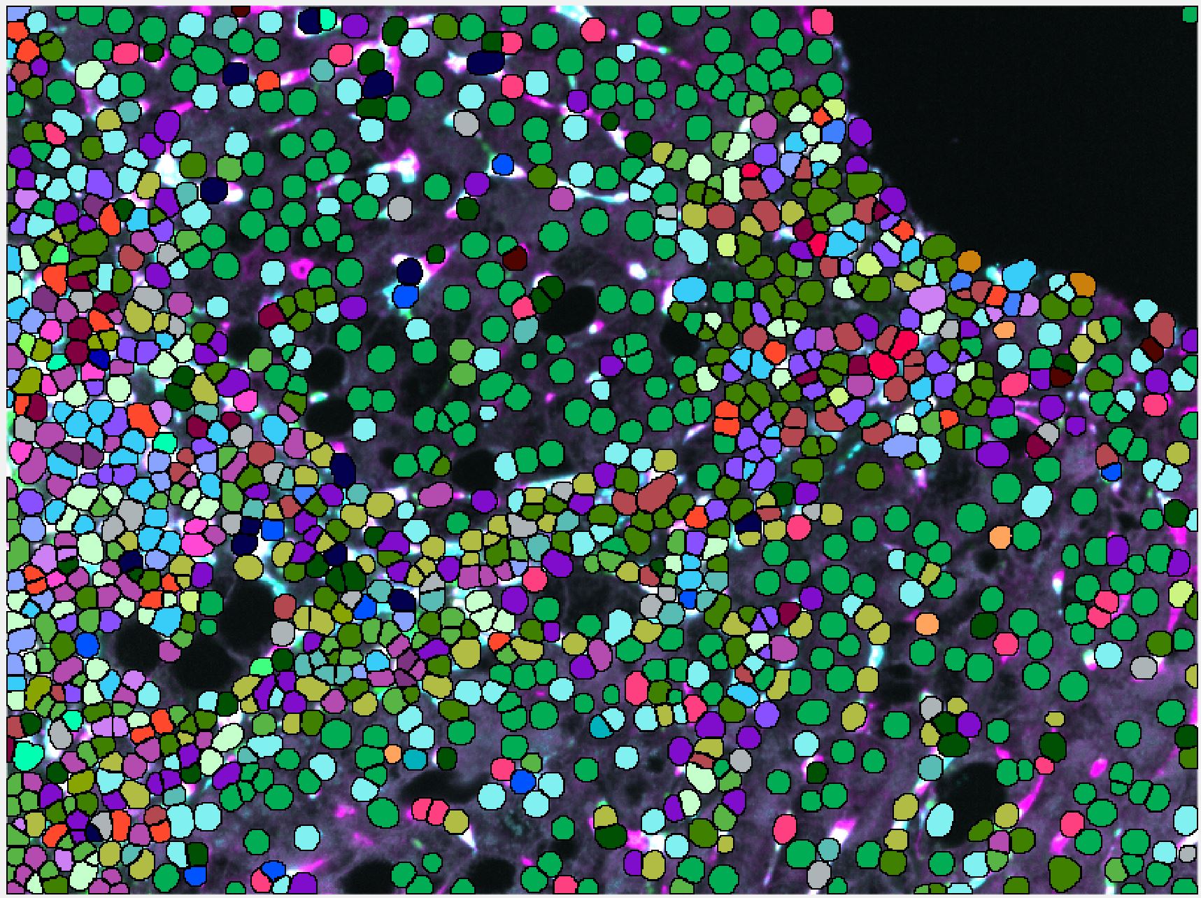
Virtual slides are subsequently analyzed in an automated fashion using the VIS software allowing us to identify, quantify, and map cell populations of interest. Specifically, the image analysis is performed using the analysis modules Tissuealign, Author, and HISTOmap. Here, we propose a strategy for the rational design of tissue multiplex assays using commercially available reagents, affordable microscopy equipment, and user-friendly software.
Using this strategy, we created one virtual slide comprising 11 biomarkers plus two frequently-used histological stains: hematoxylin and eosin (H&E) and picrosirius red (PSR). Multiple immune cell populations were identified, located, and quantified in different tissue compartments and their spatial distribution resolved using tissue heatmaps. This strategy maximizes the information that can be gained from limited clinical specimens and is applicable to formalin-fixed paraffin-embedded (FFPE) archived tissue samples, including whole tissue, core needle biopsies, and tissue microarrays. We propose this methodology as a useful guide for designing custom assays for identification, quantification, and mapping of immune cell populations in the TME.
Register for this webinar to learn about integrated multiplexing techniques for spatial resolution of immune cells in tissue sections.
Speaker

Manuel Flores, PhD candidate, Dr. Naglaa Shoukry Laboratory, CRCHUM, Université de Montréal
Manuel Flores obtained his biochemistry degree from Havana University, Cuba. In 2015, Manuel enrolled in the Immunology and Virology Master Program at Université de Montréal (Canada) and fast tracked to the Immunology and Virology PhD Program in 2016. His doctoral research project focuses on characterizing the liver resident and infiltrating immune cell populations and their role in the pathogenesis of chronic liver diseases due to persistent viral and toxic injuries, including fibrosis and hepatocellular carcinoma.
Manuel’s research interests center around the spatial organization of immune cells in the hepatic tissue microenvironment, and the delineation of the multiple cell-cell interactions and their respective biological significance in health and disease. Manuel Flores is the recipient of doctoral scholarships from University of Montreal and from Fonds de recherche du Québec – Santé (FRQS).
Who Should Attend?
- Research Scientists
- Imaging Specialists
What You Will Learn
In this webinar, participants will learn about:
- How integration of serial imaging, sequential labeling, and image alignment in the experimental design of imaging assays can greatly increase the number of markers that can be visualized simultaneously, expand the possibilities of the analysis, and extract more information from precious clinical specimens
- That virtual multiplexing determines how markers visualized in one section spatially relate to markers visualized in another contiguous section
- How the use of whole tissue sections instead of selected fields of view for the analysis results in an unbiased representation of the TME
- How the use of tissue heatmaps greatly simplify the visual representation of the spatial organization of cells in the tissue
Xtalks Partner
Visiopharm
Visiopharm® is a world leader in AI-driven digital precision pathology software. Leading biopharmaceutical companies, contract research organizations (CRO), academic medical centers, and diagnostic pathology labs all over the world use Visiopharm’s technology for tissue-based research and diagnostics. Its solutions use the latest advancements in artificial intelligence and deep learning to make the most comprehensive, highly configurable, and accurate tissue mining tools available on the market today. Visiopharm was founded in 2001 and is privately owned. The company operates internationally with over 900 licenses in more than 38 countries. Company headquarters are in Denmark’s Medicon Valley, with further offices in London, England, Munich, Germany, and Westminster, Colorado.
Related Webinars
You Must Login To Register for this Free Webinar
Already have an account? LOGIN HERE. If you don’t have an account you need to create a free account.
Create Account