
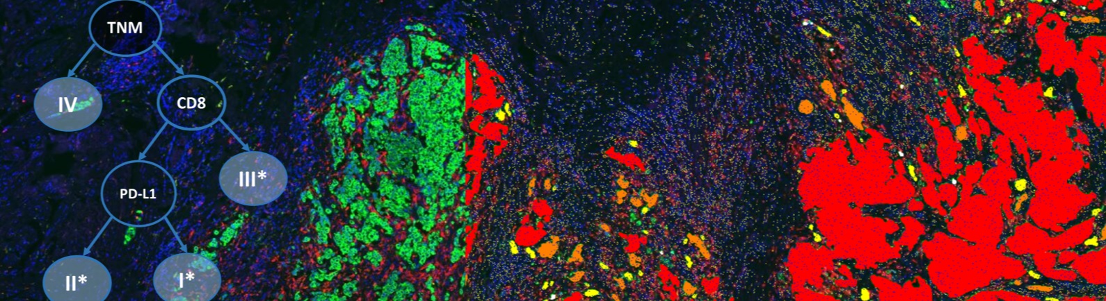
The advance of digital pathology and the availability of large-scale quantitative image analysis of highly multiplexed panels of tissue sections enable the application of big data methods in biological and clinical research. By mining multi-parametric image analysis results for statistically significant morphological and functional spatial patterns (tissue phenes) and by correlating such phenes with disease progression, new tissue-based diagnostic algorithms are being discovered.
In this webinar, Dr.Harder will present the Tissue Phenomics methodology to discover new tissue-based diagnostics using image and clinical data from prostate cancer patients with similar pathologies and known clinical outcome.
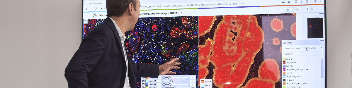
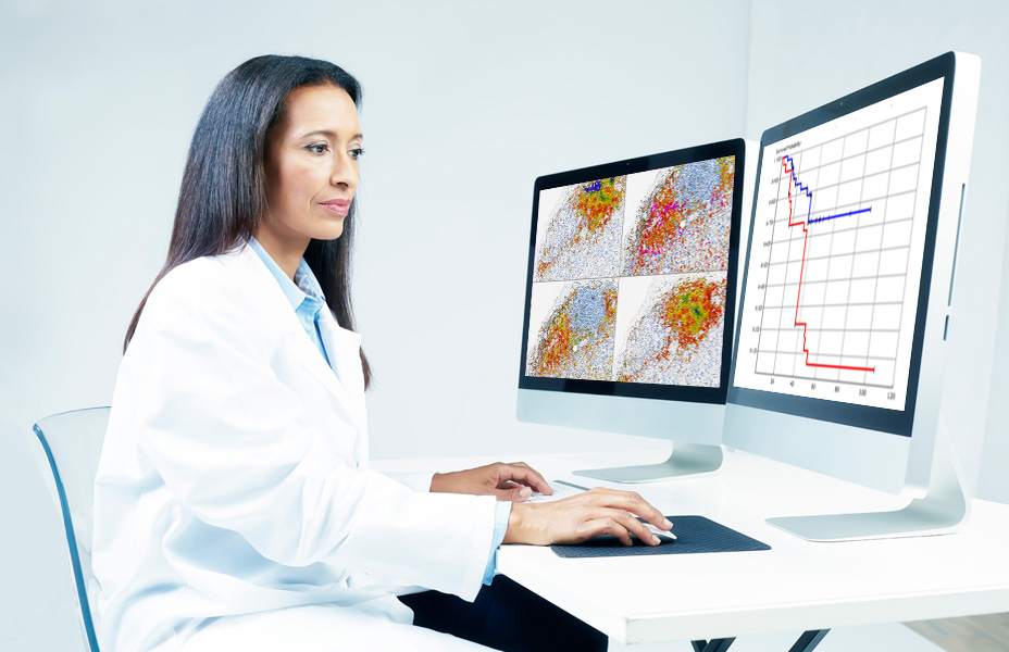
Dr. Harder will demonstrate how immunohistochemically stained tissue sections are used for:
- Extracting large amounts of image-based features from different levels of the object resolution using automatic image analysis methods.
- Discovering the most relevant image-based features by aggregating and mining for correlations with clinical data using machine learning.
- Employing relevant image-based features for the development of tissue-based prognostic and predictive biomarkers concerning individual patient clinical outcome.
In conclusion, viewers will see how Tissue Phenomics is opening the door for a new generation of diagnostic tests for the benefit of the patient.
The presented work is part of a joint project of Definiens Research with the Urological Clinic (Dr. A. Buchner, Prof. C.G. Stief) and the Institute of Pathology (Dr. H. Hessel, Prof. T. Kirchner) of the Ludwig-Maximilian-University, Munich.
Speaker

Nathalie Harder, PhD, Research Scientist, Definiens AG
Dr. Harder earned her PhD degree in Computer Science from the University of Heidelberg, Germany. Between 2004 and 2014 she was a research associate and postdoc at the German Cancer Research Center (DKFZ) Heidelberg and the University of Heidelberg in the field of Biomedical Computer Vision, focusing on the automatic analysis of live cell microscopy images (i.e. segmentation, tracking, and classification of cells and sub cellular structures). Since Jan. 2015 she is with Definiens AG, Munich, working as a research scientist on automatic image analysis and data mining of histopathological images, advancing the Tissue Phenomics approach.
Who Should Attend?
This webinar will be ideal for Medical, Pharmaceutical, Biotech and Diagnostics executives from directors and vice presidents of therapeutic areas and Chief Scientific, Medical and Executive Officers. It will also be informative for lab and study directors, and medical directors.
Xtalks Partner

Definiens
Definiens is the pioneering provider of Tissue Phenomics® solutions for biomarker and companion diagnostics development and commercialization. Definiens’ technology empowers smarter tissue-based diagnostics by leveraging quantitative tissue readouts and other big data sources. By enabling the development of powerful and precise assays for patient stratification and clinical trial enrollment, Definiens aims to dramatically improve patient outcomes. Definiens’ Tissue Phenomics® approach was awarded the 2013 Frost and Sullivan Company of the Year Award for Global Tissue Diagnostics and Pathology Imaging. For more information, please visit: http://www.definiens.com/.
Media Partner
Related Webinars
You Must Login To Register for this Free Webinar
Already have an account? LOGIN HERE. If you don’t have an account you need to create a free account.
Create Account