In this presentation, we will show examples of how histopathologic multiplexed immunohistochemistry (IHC) benefits from artificial intelligence and is used to a) gain a comprehensive understanding of Tumor Microenvironment (TME) heterogeneity, b) enhance current predictive/prognostic patient stratification and c) facilitate the development of novel biomarker signatures.
Effective response to immunotherapy relies on dynamic interactions between tumor and immune cells in the tumor microenvironment. A critical step towards improving current therapeutic strategies in immuno-oncology (IO) is to dissect the immune response to cancer and to study the composition and heterogeneity of the TME.
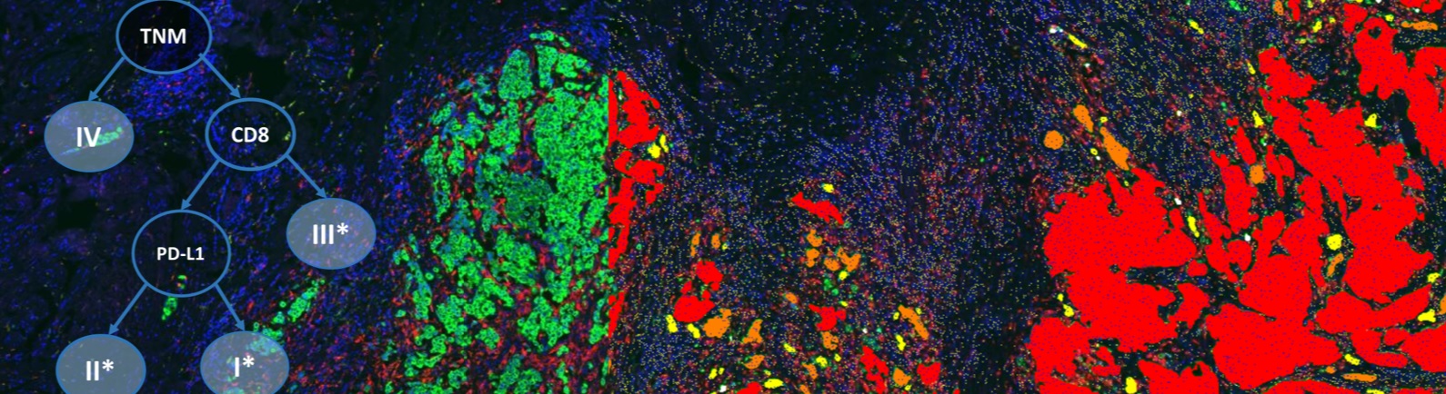
Single biomarkers, as PD-L1 IHC tests, have improved our understanding and prediction of patient’s immune response, but they are still limited in generating insights. Although PD-L1 expression can be used to predict a more favorable response to IO treatment, a significant percentage of patients with high expression of PD-L1 still do not respond. Hence, the question is whether a combination of markers can improve prediction and patient stratification. In this webinar, we show how we used automated image analysis on NSCLC biopsies to improve prediction of response to anti-PD-L1 therapy using a combination of PD-L1 and CD8.
Having shown that a combination of two markers improve prediction, we will in the second part of the webinar focus on complex patterns and demonstrate how a set of markers can be used to group patients based on biomarker profiles. The first example will show how machine learning guided feature selection from seven biomarkers improved the recurrence prediction in Prostate Cancer patients with intermediate Gleason Scores (Gleason Score: 6 – 9).
The second example will demonstrate a study using seven multiplexed immune markers to segregate NSCLC patients into six categories. The Definiens standardized marker panel (IO-Panel) consist of a combination of validated multiplex IHC assays. Based on an optimized image and data analysis protocol, the IO-Panel is designed to stratify patients into knowledge-based immune categories ranging from hot, intermediate to cold.
Identification of tissue based spatial patterns supports biological hypothesis generation and further helps us to improve our understanding of the complex immune responses to cancer. In the last part of this webinar, we will focus on the quantification of tumor heterogeneity and show how to apply artificial intelligence to identify global and local TME patterns and to profile patients based on these spatial features.
Join Ingrid Braenne in this webinar to learn and discuss how artificial intelligence and multiplexing of biomarkers can be used to extract biological insights from digital histopathology slides. Gain insights on how to profile patients based on the TME and thereby unlock a comprehensive view on the tumor immune heterogeneity. Learn how to improve the development of predictive biomarkers to effectively aid your clinical development in oncology.
Speaker

Ingrid Braenne, Ph.D., Team Manager Exploratory Data Science, Definiens AG
Dr. Ingrid Braenne received her Ph.D. in Bioinformatics from the University of Lübeck (Germany), where she applied machine learning approaches to group patients based on their genomic profiles. She has published over 20 papers and has many years of experience in multi-omics data analysis to identify the mechanisms underlying complex diseases. In her role as a senior scientist at the University of Virginia’s Center for Publish Health Genomics, she established pipelines to analyze longitudinal transcriptomics data using co-expression network analyses. Now as the team leader of Definiens’ exploratory data science group, she brings her expertise in machine learning and multi-omics data integration to the immuno-oncology field. Her team focuses on the identification of novel biomarkers and immune profiling using IHC and IF images, as well as on the integration of multi-omics data for a more detailed analysis of the tumor microenvironment.
Who Should Attend?
This webinar will be suitable for:
- Translational Scientists
- VPs of Oncology
- Directors of Immuno-Oncology programs
- Directors of Companion Diagnostics
- Directors of Profiling
- Clinical Trial Managers/Directors/VP
What You Will Learn
In this webinar, participants will explore:
- The strengths of multi-biomarker analysis combined with AI
- Comprehensive immune profiling using multiplexed markers
- How to achieve better patient stratification using IHC multiplex imaging analysis and genomic information
Xtalks Partner

Definiens
We improve patient lives by unlocking the tissue phenome.
In oncology, therapeutic strategies have shifted from a direct assault on cancer cells to recruiting the immune system for that purpose. Our mission is to accelerate breakthroughs for this approach by helping scientists leverage Tissue Phenomics to deepen understanding of disease biology and immune system mechanisms, to bring multi-omics data into a cancer-relevant context, and to facilitate the translation of new insights into novel therapies and treatment strategies. Our vision is to create unique patient profiles for an individualized standard of care, where patients experience fewer side effects and live longer.
Definiens’ Tissue Phenomics approach was awarded the 2013 Frost and Sullivan Company of the Year Award for Global Tissue Diagnostics and Pathology Imaging. For more information, please visit: www.definiens.com.
Related Webinars
You Must Login To Register for this Free Webinar
Already have an account? LOGIN HERE. If you don’t have an account you need to create a free account.
Create Account