In translational oncology research, understanding and evaluating the phenotypic profiles of cells in the tumor immune microenvironment can yield a deeper understanding of the complex interactions within the immunological landscape of the tumor.
Multiplex immunofluorescence can study this interplay through rapid identification, quantification and mapping of many cell types. Having the flexibility to look for specific biological markers and their co-localization within this environment allows researchers to better understand the mechanisms of action for compounds of interest.
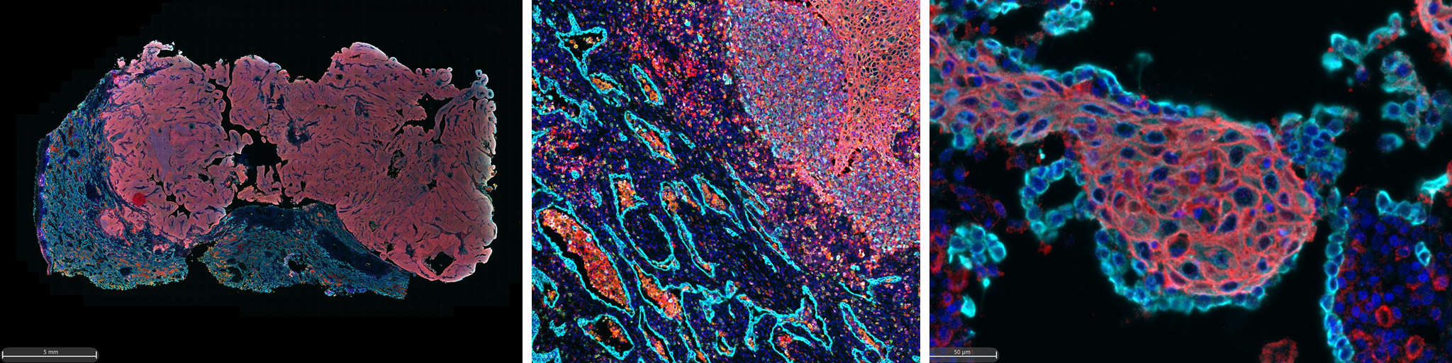
Here, Scott Lawrence shows his work at the NCI using a customized 8-plex mIF panel to study markers in a multitude of cancer types and discusses the insights gained from data gathered through staining, imaging and analysis.
Speakers

Scott Lawrence, (M.S., HT(ASCP)) [C] Associate Scientist, Cancer Genomics Research Laboratory (CGR) Division of Cancer Epidemiology and Genetics, NCI, FNLCR, Leidos Biomedical Research
Scott Lawrence is an associate scientist in the Molecular and Digital Pathology Laboratory for Leidos Biomedical Inc. The group primarily supports the Division of Cancer Epidemiology and Genetics (DCEG) of the NCI with all histopathology applications from tissue preparation though staining, imaging, and analysis. He started his career in a veterinary histopathology lab where he established digital imaging and analysis applications for preclinical work in mice and rats. Afterwards he worked with a pharmacodynamics group developing slide-based assays for phase 0 clinical trials focusing on standardizing multiplex immunofluorescent acquisition and analysis of tissue and circulating tumor cells. Lawrence has worked in the field for over 17 years bridging experience from the histology wet lab with digital imaging, automation and analysis.

Karan Sharma, Associate Director, Product Portfolio, Ultivue
Karan Sharma is currently the Associate Director of Product Management for the Ultivue portfolio and is based in Cambridge, Massachusetts. Prior to joining Ultivue, Sharma was the Global Product Manager for the RabMAb antibody portfolio at Abcam where he focused on the development of markers for immuno-oncology, neuroscience and pre-clinical mouse research. He has a successful track record of about bringing new antibody-based solutions to market in the life sciences industry and is passionate about all things multiplex.
Who Should Attend?
For researchers in the translational oncology space looking to better understand the clinical utility of multi-parameter biomarker signatures to characterize the tumor immune microenvironment.
What You Will Learn
- Multiplex immunofluorescence is a beneficial tool in understanding and characterizing the tumor microenvironment
- InSituPlex is a ready-to-deploy, high-throughput mIF platform that is workflow-agnostic and provides whole-slide biomarker profiling
- Fast, customizable mIF panels accelerate the hypothesis-to-answer workflow and can be key in gaining insights about targets of interest tailored to specific projects
Xtalks Partner
Ultivue
Ultivue provides researchers with multiplex biomarker assays for tissue phenotyping and digital pathology. Ultivue’s InSituPlex® technology enables scientists to unmask and analyze the true biological context of tissue samples. With the ability to run a same-slide fluorescent and H&E stain, Ultivue’s technology is enabling pathologists to connect traditional morphological analysis with multiplexed immunofluorescent data for comprehensive single-cell phenotyping.
Related Webinars
Profiling Cancer Biology Using Spatial Phenomics
You Must Login To Register for this Free Webinar
Already have an account? LOGIN HERE. If you don’t have an account you need to create a free account.
Create Account