
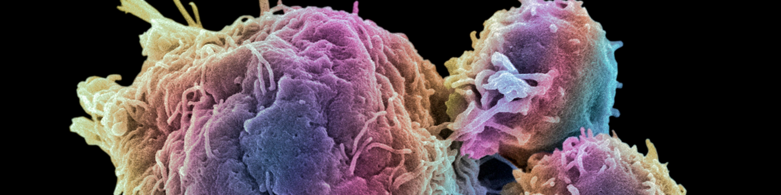
Despite the effectiveness of anti-retroviral therapy (ART) in significantly improving health, quality of life and reducing mortality among HIV-positive individuals, new research shows that persistent infections may be due to the ability of the virus to take refuge and hide in distinct subsets of immune T cells. This may help explain why HIV is adequately controlled, but not eradicated, with current treatments. The identification of T cells that ‘hide’ the virus has significant implications for developing therapeutic strategies to improve the treatment and long-term management of HIV.
Xtalks spoke to lead author on the research, Mohamed Abdel-Mohsen, PhD, assistant professor, Vaccine & Immunotherapy Center at the Wistar Institute, about the emerging utility of identifying glycomic, or sugar, signatures on the surface of immune cells to help in their identification and potential targeting. His research team’s study has been published in Cell Reports.
Dr. Abdel-Mohsen was particularly interested in studying the cell surface glycome of HIV-infected cells because, “In the HIV field, there was nothing about [cell surface glycomes] at all. However, there is a lot of interest in trying to identify any feature of HIV-infected cells that can differentiate them from uninfected cells. On one hand, those features can be used to target those cells, but also, they will help us learn a lot about the biology of HIV persistence.”
Dr. Abdel-Mohsen and his group at the Wistar Institute, a non-profit biomedical research center located on the campus of the University of Pennsylvania, studied mechanisms of persistent HIV infection and discovered that there are hidden reservoirs of HIV in immune T cells. They found that by specifically hiding in a rare population of CD4 positive T cells, the virus is able to evade therapy and persist in the body. Identification of these viral reservoirs through specific markers could therefore be an important strategy in eradicating infection.
The results of persistent, underlying HIV infection include heightened immune activation and inflammation (leading to a number of diseases), ongoing damage to multiple organ systems and reduced life expectancy.
Related: FDA Approves Rukobia for HIV Patients with Limited Treatment Options
ART effectively halts disease progression by inhibiting viral replication in host immune cells, however, even after therapy, low, residual amounts of virus remain in the blood and tissues.
Researchers have known for a while that viral eradication during therapy is hindered by the ability of HIV to maintain persistent infection primarily in CD4+ T cells, and in other types of cells in the blood as well. Studies have identified the presence of two types of HIV-infected CD4+ T cells:
- ‘Transcriptionally inactive’ cells that do not typically produce viral RNA or viral proteins
- ‘Transcriptionally active’ cells in which HIV RNA is actively transcribed to make copies of the virus (despite long-term ART)
While latent HIV infection has been thought to largely reside in the inactive pool of HIV-infected cells, the new study builds on recent research that shows the presence of a significant virus-harbouring, transcriptionally active CD4+ T cell pool, and that this pool can be identified by cell surface-specific markers.
T Cell Glycomic Signatures in HIV
Unique and diverse patterns of cell surface protein glycosylation (the addition of sugar molecules to proteins) make up specific glycoimmune profiles that have key functional roles in regulating cellular processes and immune functions. Moreover, altered glycan structures can serve as biomarkers for cancer and infectious diseases, according to the research paper.
Dr. Abdel-Mohsen told Xtalks that the cancer field has greatly informed our current knowledge of cell surface glycomes. In particular, the surface of cancer cells is covered in sugars including the sugar fucose, which binds to a sugar-binding protein called selectin that exists on endothelial cells, allowing them to move. In addition, it has recently been found that cancer cells also cover themselves with a sugar called sialic acid, which binds to sialic acid-binding proteins called sialic acid-binding immunoglobulin-like lectin (Siglecs).
This interaction allows cancer cells to evade immune surveillance as Siglecs are expressed by many immune cells including NK cells. The binding of sialic acid on the surface of cancer cells to NK cells sends a very strong inhibitory signal to the NK cells, allowing the cancer cells to show a fake ‘self’ to the immune cells and escape their action, explained Dr. Abdel-Mohsen.
Given the importance of cell surface sugars in immune cell interactions, Dr. Abdel-Mohsen and colleagues worked to characterize the cell-surface glycome of HIV-infected cells.
In the study, the researchers conducted a comprehensive glycomic analysis of HIV-infected cells isolated from a primary cell model of HIV latency. They found that the cell surface of HIV-infected transcriptionally active CD4+ T cells had high levels of fucosylated carbohydrate ligands compared with HIV-infected transcriptionally inactive cells. Fucosylation is the attachment of a specific sugar molecule called fucose to proteins on the surface of cells. Fucosylation has been shown to be critical for T cell activation. These findings were confirmed in CD4+ T cells obtained from HIV-infected individuals on ART.
In addition, the research team found that Sialyl-LewisX (SLeX), a fucosylated antigen that functions as a cell extravasation mediator, is highly expressed on the surface of HIV-infected transcriptionally active cells in vivo. It was observed that active HIV transcription, but not cellular activation, induces SLeX cell-surface expression in vitro. Moreover, cells with high levels of SLeX were found to be enriched with markers associated with HIV susceptibility, signaling pathways that drive HIV transcription, as well as pathways involved in leukocyte extravasation and T cell trafficking.
High SLeX levels are in fact a key feature of some metastatic cancer cells. The enrichment of cellular trafficking pathways in transcriptionally active T cells could thus play a role in the persistence of HIV in tissues, which are the sites where HIV primarily ‘hides’ during ART.
Dr. Abdel-Mohsen explained, “We zoomed in to try to find the glyco-antigen – because sugars are [located within] a glycoprotein – and found it’s actually in a particular glyco-antigen called SLeX, which is highly expressed in many cancer cells, allowing them to metastasize. In our work, [it] might allow those cells that are expressing HIV RNA, and have high level of SLeX to migrate more from blood and tissues, which will allow them to hide.”
“On the other hand, we found that cells that are infected with HIV but do not make RNA (transcriptionally inactive) have lower than normal levels of SLeX. So, we identified the feature that differentiates these two cells from normal, from each other and that might have an implication in trafficking of these cells between blood and tissues,” he said.
HIV: An Infection of Tissues
HIV is typically seen as an infection circulating in the blood. However, Dr. Abdel-Mohsen said that, “In the last few years, it has become very clear that HIV is an infection of tissues because that is where HIV hides during ART. It’s like HIV uses blood similar to how cars use highways: eventually they will go to cities and live there, but they use highways just for trafficking and that’s what HIV does. Ninety-nine percent of HIV in the body is actually in the tissues.”
Tissues serve as sanctuary sites for HIV, explained Dr. Abdel-Mohsen. This is because tissues may not be optimally penetrated by ART, and may be ‘immune privileged’ sites as they lack cytotoxic CD8 T cells, and therefore do not have optimal immune function. “So usually that’s why HIV lives in the tissues. There is a lot of interest recently in how they hide in tissue, and how they move in and out of tissue. And because we could confirm this feature of high level of fucose, [we think] it may play a role,” said Dr. Abdel-Mohsen.
Sugar-Based Signatures to Identify HIV-Infected Cells
The distinct glycosylation patterns could serve as a distinguishing feature between transcriptionally active and transcriptionally inactive HIV-infected CD4+ T cells, as they were not found on the surface of the latter. Interestingly, the study also found that HIV could promote these cell-surface glycomic changes itself.
“With recent advances that we are making in the fields of glycobiology and glycoimmunology, it has become clear that the sugar molecules present on the surface of immune cells play a critical role in regulating their functions and fate,” said Dr. Abdel-Mohsen.
“However, the relevance of host cell-surface glycosylation in HIV persistence remained largely unexplored, making it a ‘dark matter’ in our understanding of HIV latency. For the first time, we described a cell-surface glycomic signature that can impact HIV persistence.”
Work from different fields, including evolutionary biology, suggest that there are four fundamental building blocks of life: DNA and RNA (nucleic acids), protein, lipids and sugars. Dr. Abdel-Mohsen explained to Xtalks that, “When these building blocks are arranged [in terms of conservation], DNA, or the genome, is the most evolutionary conserved and the least diverse, and then it’s the transcriptomes (RNA), the proteome, lipidome and then the glycome, which is the least evolutionary conserved, making it the most rich and diverse in information.” He said if you want to try to find a biomarker, fingerprint or blueprint of a process, you most likely will find a fingerprint at the glycan level, compared to lipids, protein, RNA and DNA.
This is why Dr. Abdel-Mohsen and his group “hypothesized that HIV infection, even if it’s latent or even during ART, it still may be making a fingerprint on the surface glycome of those cells, similar to what’s going on with cancer. We were hoping that by identifying [it], maybe it could teach us something about HIV-infected cells.”
These findings can help identify specific subsets of virally active immune cells based on their specific cell surface glycosylation profiles. Importantly, carbohydrate-based therapeutic vaccines can be designed using the unique glycan profiles of these active HIV-infected cells, providing a potential step closer to eradicating and curing the disease.
The importance of identifying and combining signatures across multiple realms, from gene expression to the proteome to the glycome, is important in the effective identification, classification and therapeutic targeting of disease in a more specified and personalized manner, and in contributing to our overall understanding of biological processes.
Dr. Abdel-Mohsen highlighted the fact that the field of glycan biology has experienced significant growth in recent years, owing to advancements in technologies for studying glycans. He is also enthusiastic about what the future holds for furthering our understanding of HIV infection in the context of glycoimmunology.
In fact, the National Research Council (US) Committee (National Academies Press; 2012) recognizes that, “Glycans are directly involved in the pathophysiology of every major disease … Additional knowledge from glycoscience will be needed to realize the goals of personalized medicine and to take advantage of the substantial investments in human genome and proteome research and its impact on human health.”


Join or login to leave a comment
JOIN LOGIN