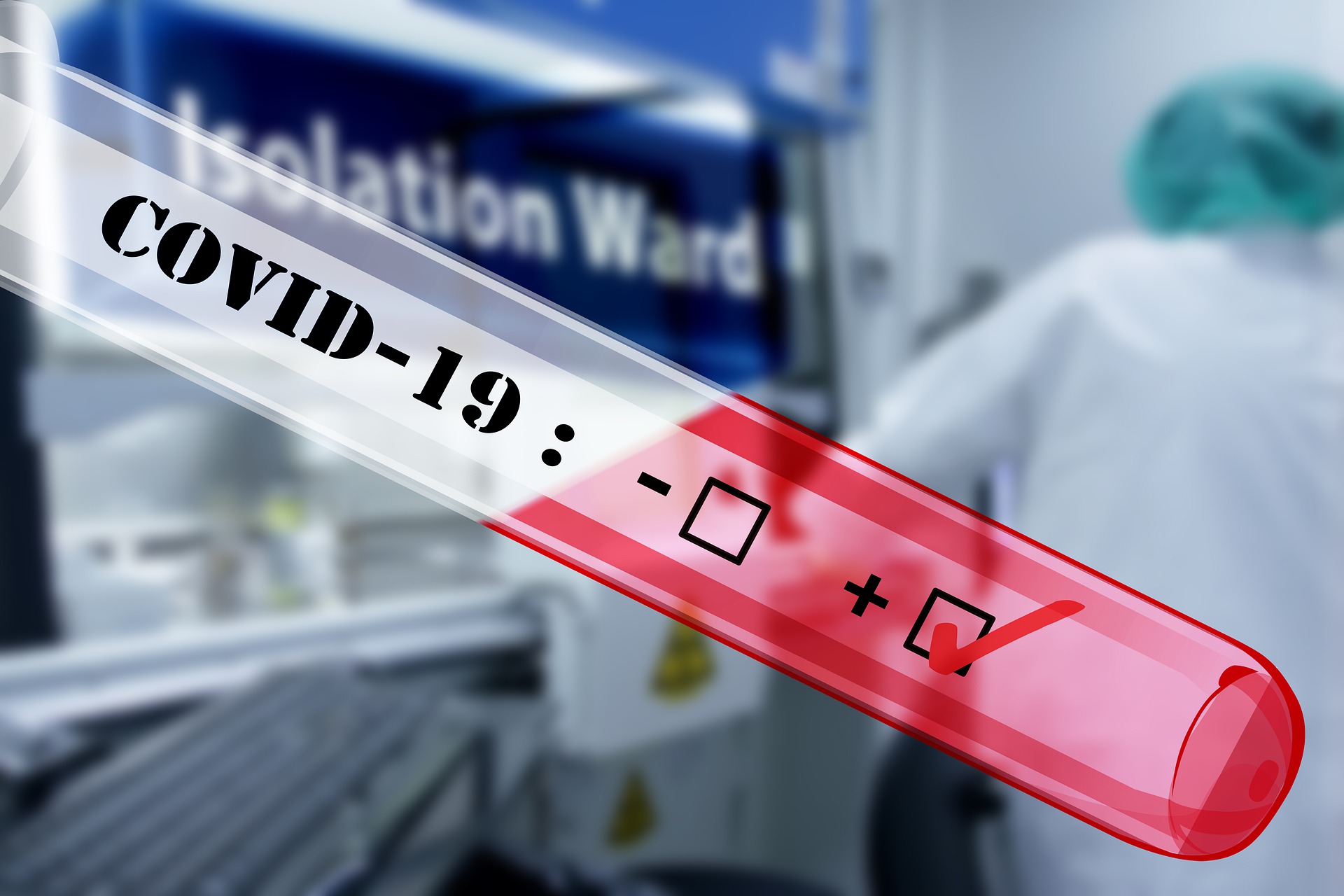
At the end of 2019, cases of pneumonia of an unknown origin were proliferating in Wuhan city, China (1). This outbreak, infecting thousands of people and killing hundreds in China, soon spread outside of the country and started infecting people all around the globe in January 2020. It was rapidly discovered that the causative agent for this disease was a new beta-coronavirus (2) resembling the severe acute respiratory syndrome coronavirus (SARS-CoV) (3). In consequence, the virus was named SARS-CoV-2 (or alternatively 2019 novel coronavirus, 2019-nCoV) and the associated disease COVID-19 by the World Health Organization (WHO). On March 25th, 2020, 414,179 cases of COVID-19 have been reported around the world, causing the death of 18,440 patients (4) and becoming a global health threat.
Coronaviruses are a wide family of enveloped, single-stranded positive-sense RNA viruses. They can infect both animals and humans, usually causing mild respiratory tract infections (5). However, some members of this family, referred as highly pathogenic human CoVs (HCoVs), have emerged from animal reservoirs in the last two decades and led to global epidemics with much higher morbidity and mortality. In 2004, the SARS-CoV provoked an epidemic in China while the Middle East respiratory syndrome coronavirus (MERS-CoV) infected thousands of people in the Eastern Mediterranean region in 2012 (6). SARS-CoV-2 is the seventh member of the coronavirus family with an ability to infect humans. Like for SARS-CoV and MERS-CoV, the SARS-CoV-2 is mainly transmitted by droplets and close contact and its associated symptoms include fever, fatigue, dry cough and pulmonary pain. It can affect all age spectrum, but the disease is generally more severe in older people, frequently having pre-disease disorders like hypertension, diabetes or chronic obstructive pulmonary disease (7).
Analysis of seven clinical samples suggested that SARS-CoV-2 shares 79.5% sequence identity with SARS-CoV (8). The receptor binding viral protein is the spike (S) protein. The S1 subunit contains the receptor binding domain (RBD) whereas the S2 subunit is responsible for membrane fusion and internalization in host cells (9). The RBD binds to the same receptor in SARS-CoV and SARS-CoV-2: the angiotensin converting enzyme II (ACE2) (10). This receptor is expressed mainly in a small subset of cells in the lung called type 2 alveolar cells (11). Even though it has been reported that SARS-CoV can directly infect macrophages and T cells (12), it is not known if SARS-CoV-2 can infect any immune cells. Structural and biophysical analysis revealed that the S protein of SARS-CoV-2 has approximately a 10- to 20-fold higher affinity for its receptor (13), potentially explaining it concomitant facility to infect human subjects.
Currently, not much is known about the innate immune status of infected patients, but lymphopenia is one of their major characteristics. An excessive inflammatory response is suggested by an increase of circulating neutrophils, reduced total lymphocytes and high levels of serum IL-6 and C-reactive protein. Cytokine profiles seem to emerge among critical patients showing higher plasma concentrations of IL-2, IL-7, IL-10, G-CSF, IP-10, MCP-1, MIP-1A and TNFα (16). These cytokines probably play a role in the pulmonary disease developed during infection. These features are once again like what is observed with SARS-CoV and MERS-CoV and underline the role of leukocyte alteration and cytokine storm in COVID- 19 pathogenesis.
T cell response in SARS-CoV was shown as mainly CD8-oriented. Virus-specific T cells from severe patients tended to be of central memory phenotype with a significantly higher frequency of polyfunctional CD4+ T cells (IFNγ, TNFα, and IL-2) and CD8+ T cells (IFNγ, TNFα and degranulated state), as compared with the mild-moderate group. Strong T cell responses correlated significantly with higher neutralizing antibody titers while more serum Th2 cytokines (IL-4, IL-5, IL-10) were detected in patients in critical condition (17). In term of specificity, 70% of responses were mounted against structural proteins (spike, envelope, membrane and nucleocapsid) (18). The first reports on anti-SARS-CoV-2 cellular response showed that infected patients had low B, T (both CD4 and CD8) and NK cell counts, increased naïve helper T cells and decreased memory helper T cells. In addition, regulatory T cells were also found as decreased in patients presenting COVID-19 (19). The decrease in T cells might be due to the elevated serum levels of TNFα, IL-6 and IL-10 negatively regulating T cell survival or proliferation (20). Immune exhaustion was also reported as a characteristic of T cells during COVID-19. Indeed, patients with severe disease showed CD8 T cells expressing higher levels of exhaustion markers such as PD-1 and Tim-3 than patients with milder pathology (20).
SARS-CoV and MERS-CoV infections induce seroconversion by 14 days after disease onset in most patients. Neutralizing antibodies were reported as long as 2 years after infection (21). On the other hand, delayed or weak antibody responses were associated with severe outcomes for both types of coronavirus infections (22). A significant part of neutralizing antibodies is directed against the S protein, rendering it a promising target for vaccine development (23). In a preliminary study, one patient infected with SARS- CoV-2 showed a peak of specific IgM 9 days after disease onset and a class switch to IgG after two weeks (24). Sera from patients with COVID-19 showed cross-reactivity with SARS-CoV but no other coronaviruses, although some unpublished reports of anti-MERS cross-reactivity exists. Neutralization of SARS-CoV was also observed by sera from all COVID-19 tested patients (8).
Vaccines and Therapeutics in development
In response to the outbreak, an unprecedent effort to develop prophylactics and therapeutics has begun. The WHO maintains a list of vaccine candidates (https://www.who.int/blueprint/priority-diseases/key- action/novel-coronavirus-landscape-ncov-21march2020.PDF?ua=1 ) as of March 21st, the number of candidates stood at 43. The participation in this effort is broad, ranging from academic institutions to the largest vaccine manufacturers in the world. The approaches are also varying from the mRNA approach put into the clinic by Moderna and NIAID to VLPs expressed in various host cells. The goal of all of these candidates will be prime the immune system via humoral and/or cell-mediated immunity to limit the virus’s ability to cause disease.
Many of the organizations also have varying levels of ability to assess the immune response to these candidate vaccines in both the time and throughput required to quickly advance these assets. As part of their R&D Blueprint (https://www.who.int/blueprint/priority-diseases/key-action/COVID-19-vaccine- trial-synposis.pdf?ua=1), the WHO identified that immune correlates will be a necessary evaluation criterion. A standardized set of assays will provide regulatory agencies with data set which will be comparable across different modalities to support licensure. These vaccines at best, may be available in >1 year, at an extremely accelerated pace.
To provide nearer term relief potentially, several therapies are also being evaluated ranging from off-label use of currently licensed products to new biologics and other modalities aimed to reduce burden of disease. Some of these include production of neutralizing antibodies targeting the COVID-19 Spike protein which would inhibit viral attachment to receptor bearing host cells. Small molecule-based antagonists to ACE2 (the ligand for viral Spike protein) are marketed for other indications and being assessed as a therapy for COVID-19. The ability of these therapies to provide neutralizing effects will also be determined via methodologies identified below. On March 24th, the FDA approved of the use of convalescent plasma to those patients that are currently battling severe disease.
Rational vaccine design
The use of world leading T cell epitope prediction tools allows the selection of epitopes and antigenic regions with a broad HLA coverage. Our specialized in vitro assays can be used for the experimental validation of the selected epitopes and constructs (mRNA, proteins, peptides, antibodies) using primary human immune cells. Analysis of the in vitro immune responses is done using multicolor flow cytometry, ELISA, Luminex and multicolor Fluorospot.
Protein Sciences
Recombinant antigens are key reagents in infectious disease research. They are widely used in the context of antibody and vaccine development. Our Protein Sciences (PS) platform enables an efficient production of recombinant proteins and antibodies against coronaviruses with a broad expertise in molecular biology, protein expression, protein purification and structural / physicochemical characterization. Our PS colleagues routinely assist the pharmaceutical industry using state-of-the-art instrumentation to produce R&D grade proteins that are rigorously characterized. These scientists solve protein expression and purification challenges and provide critical components for multiple immunoassays in anti-SARS- CoV-2 research.
Like other coronavirus, the SARS-CoV-2 strain encodes two key target proteins, the spike glycoprotein and the nucleocapsid protein. The spike protein (S-protein) is composed of two subunits, S1 and S2. The S1 subunit contains the receptor binding domain (RBD) and the S2 contains elements required for the membrane fusion. The nucleocapsid protein (N-protein) is very conserved, has a structural role and is involved in RNA synthesis. Different recombinant forms of these proteins are now available in companies such as Nexelis to aid the efforts of developing vaccines against this virus. Additional coronavirus proteins can also be produced in addition to the S- and N-protein to help advance research projects based on a different pathway.
Preclinical services
Nexelis has different tools that could be used to advance candidates in the preclinical phase. Immunogenicity studies could be performed in various species such as ferret and non-human primates (NHP). Flow cytometry in NHP is as flexible as in humans, however in ferrets it’s generally limited to classical population markers and cytokines. Furthermore, we have already developed models relevant to study several key mechanisms found in COVID-19 such as LCMV Clone 13 chronic infection in mouse for exhaustion monitoring and RSV infection in cotton rats or mouse for exacerbated lung pathology.
In addition, antisera can be produced in different species to be used as controls in in vitro assays, such as ferret which is currently developed. In term of analysis, we plan to develop antigen specific ELISAs, flow cytometry measurement of T cell parameters and neutralization assays using pseudo virions for the three most relevant animal models and human. In general, we also have the capacity to measure both the innate (ELISA, Multiplex) humoral (ELISA, ADA, neutralization assays with pseudo virions) and cellular (Flow Cytometry, ELISPOT) immune responses. In vivo imaging for mRNA vaccines is also a development step that we could perform.
As non-regulated laboratory, we have the possibility to start assays development and preliminary results in humans both for humoral and cellular immunity.
Assay Development
Based on what is known on the cellular response to SARS-CoV and SARS-CoV-2, it represents an important goal for vaccines and therapeutics to stimulate CD4 and CD8 T cells antiviral responses. Different assays will be developed by Nexelis to ensure coverage of all the important aspects of these responses.
First, the ability of CD4 and CD8 T cells to respond to antigens following vaccination will be assessed by Flow Cytometry. The cocktail used will include lineage classical markers (CD45, CD3, CD4, CD8) but also surface molecules assessing activation and proliferative status (CD62L, HLA-DR, CXCR5, Ki67, etc.) and of course intracellular cytokine to assess antiviral functionality (IFNγ, TNFα, IL-2). Since it was demonstrated in literature that Th2 cytokines (IL-4, IL-5, etc.) could lead to exacerbated immunopathology (similar to the one observed with FI-RSV vaccination), it will be important to include them also to ensure their absence and therefore candidates’ safety. In addition, the cytotoxic potential of CD8 T cells will be evaluated by measuring degranulation molecules like perforin/granzyme B. All these factors will be available for measurement following stimulation with the different SARS-CoV-2 antigens generated by the Protein Sciences department (Spike protein, RBD, Nucleocapsid). In parallel, individual antiviral factors such as IFNγ could also be measured upon ex vivo re-stimulation with the same antigens using ELISPOT assays while Fluorospot can be used for simultaneous detection of multiple analytes to document the breadth of the immune response, especially when cell numbers are limited.
It was also demonstrated that subpopulation of CD4 and CD8 T cells could be unbalanced in a context of SARS-CoV-2 infection. In consequence, evaluating the capacity of candidates, particularly immunotherapies to reverse this phenotype, represents a major readout. For that, a subpopulation phenotyping cocktail will be composed of lineage markers (CD45, CD3, CD4, CD8) and subpopulation markers (CD45RA, CD45RO, CD28, CD127, CCR7, HLA-DR, CD62L).
Another aspect of the T cell response is the exhaustion phenotype that was denoted in patients presenting the most severe cases of COVID-19. In a context of efficacy studies or clinical testing, we’ll therefore develop a phenotyping cocktail that will include lineage markers (CD45, CD3, CD4, CD8) but also standard immune checkpoints frequently associated with exhausted phenotypes (PD-1, LAG-3, Tim-3, etc.). This approach will particularly be valuable for the evaluation of immunotherapies aiming to disrupt shutdown signals.
Via our immuno-oncology specialized services, we offer a suite of immune potency assays to screen compounds that reverse T cell exhaustion and inhibit immune check points (T cell exhaustion assays Mixed Lymphocyte Reaction Assays, CMV/SEB activation assays).
High-throughput testing
The facilities at Nexelis, based in previous history as part of large pharmaceutical organization has enabled equipment and practices to support high-throughput testing to meet rapid turn around time requests and handle the capacity to support this unprecedented demand. Automated instrumentation for assay performance and analysis also serves to reduce variability between runs which will be critical during the clinical evaluations. Novel technologies for analysis of viral neutralization assays increase the number of samples an analyst can test in a week by orders of magnitude. As potentially a number of the candidates enter larger Phase IIb or III clinical studies, the ability to analyze large numbers of samples within a condensed timeframe will be critical to the successful licensure of these investigation prophylactic or therapeutic strategies.
Public Health England partnership
Nexelis has established a strategic partnership with Public Health England’s (PHE) National Infection Service to engage in the improvement of global human health. This partnership is focused on vaccine and antiviral projects with the objective of jointly developing new pre-clinical and clinical assays to support R&D and clinical biopharmaceutical teams.
As such, PHE’s Vaccine Research Groups led by Dr Bassam Hallis have developed animal models (non- human primates and ferrets) using wild-type SARS-CoV-2 strain and various tools such as anti- SARS-CoV-2 spike protein IgG ELISA, viral neutralizing assay, diagnostic RT-qPCR, pseudovirus neutralizing assay and cellular immunology assays to support global epidemiological studies and clinical trials for vaccines including two programs funded by CEPI and two by FDA. Several monoclonal antibodies and anti-viral compounds are also being analyzed.
The need for an immunology toolbox SARS-CoV-2
Working with a contract organization equipped to run immunogenicity-related projects from the lead screening stage until phase III provides the pharmaceutical company developing a drug or vaccine with the confidence that expertise developed within the team is not lost in between two stages of development and translational. We believe the fluid pipeline of our services to be a determinant reason of our expedited turnaround times.
In the area discussed hereto, however, none of the outsourcing companies with vaccine R&D and testing capabilities already have at their disposal qualified assays covering much of the potential demand of the industry in the humoral and cellular areas.
Subject to appropriate funding conditions, we would suggest to proactively develop and qualify a somehow exhaustive panel of immunoassays made available to vaccine developers saving a minimum of 3 months in development time and potentially making a vaccine available 3 months earlier.
It is not yet known if the current event will be a single pandemic or if we need to be prepared to also have yearly seasonal COVID-19 events. Should it be the case, the work done would also enable us to test the efficacy of seasonal vaccine candidates that would be needed to protect the population in 90 to 120 days as it is done with seasonal flu vaccines.
Learn more with Nexelis in an upcoming webinar titled The SARS-CoV-2 Toolbox: Innovative Initiatives to Support the Development of Vaccines & Therapeutics.
This article was created in collaboration with the sponsoring company and the Xtalks editorial team.
1.Report of clustering pneumonia of unknown etiology in Wuhan City. Wuhan Municipal Health
Commission. 2019:(http://wjw.wuhan.gov.cn/front/web/showDetail/2019123108989).
2. Lu R, Zhao X, Li J, et al. Genomic characterization and epidemiology of 2019 novel coronavirus:
implications of virus origins and receptor binding. Lancet. 2020; doi:10.1016/S0140-
6736(20)30251-8.
3. Zhu N, Zhang D, Wang W, et al. A novel coronavirus from patients with pneumonia in China,
2019. N Engl J Med. 2020; doi: 10.1056/NEJMoa2001017.
4. World Health Organization situation report 65 of COVID-19.
5. Zumla A, Chan JF, Azhar EI, Hui DS, Yuen KY. Coronaviruses – drug discovery and therapeutic
options. Nat Rev Drug Discov. 2016;15(5):327-347.
6. Paules, C. I., Marston, H. D. & Fauci, A. S. Coronavirus infections—more than just the common
cold. JAMA 323, 707–708 (2020).
7. Guan W et al. Clinical characteristics of 2019 coronavirus infection in China. MedRxiv 2020.
doi.org/10.1101/2020.02.06.20020974.
8. Zhou P, Yang XL, Wang XG, et al. A pneumonia outbreak associated with a new coronavirus of
probable bat origin. Nature. 2020.
9. He Y, Zhou Y, Liu S, et al. Receptor-binding domain of SARS-CoV spike protein induces highly
potent neutralizing antibodies: implication for developing subunit vaccine. Biochem Biophys Res Commun. 2004;324(2):773-781.
10. Li W, Moore MJ, Vasilieva N, et al. Angiotensin-converting enzyme 2 is a functional receptor for
the SARS coronavirus. Nature. 2003;426(6965):450-454.
11. Zhu N, Zhang D, Wang W, Li X, Yang B, Song J, et al. A Novel Coronavirus from Patients with
Pneumonia in China, 2019. N Engl J Med. 2020; 382(8):727-33.
12. Perlman S, Dandekar AA. Immunopathogenesis of coronavirus infections: implications for SARS.
Nat Rev Immunol. 2005;5(12):917-27.
13. Wrapp D, Wang N, Corbett KS, et al. Cryo-EM structure of the 2019-nCoV spike in the prefusion
conformation. Science. 2020.
14. Wu Z, McGoogan JM. Characteristics of and Important Lessons From the Coronavirus Disease
2019 (COVID-19) Outbreak in China: Summary of a Report of 72314 Cases From the Chinese
Center for Disease Control and Prevention. JAMA. 2020.
15. Hui DSC, Zumla A. Severe Acute Respiratory Syndrome: Historical, Epidemiologic, and Clinical
Features. Infect Dis Clin North Am. 2019;33(4):869-889.
16. Huang C, Wang Y, Li X, et al. Clinical features of patients infected with 2019 novel coronavirus in
Wuhan, China. Lancet. 2020;395(10223):497-506.
17. Li CK, Wu H, Yan H, Ma S, Wang L, Zhang M, et al. T cell responses to whole SARS coronavirus
in humans. J Immunol. 2008;181(8):5490-500.
18. Shin HS, Kim Y, Kim G, Lee JY, Jeong I, Joh JS, et al. Immune Responses to Middle East
Respiratory Syndrome Coronavirus During the Acute and Convalescent Phases of Human
Infection. Clin Infect Dis. 2019;68(6):984-92.
19. Qin C, Zhou L, Hu Z, Zhang S, Yang S, Tao Y, Xie C, Ma K, Shang K, et al. Dysregulation of immune
response in patients with COVID-19 in Wuhan, China. Clin Infect Dis, published ahead of print
March 12, 2020. https://doi.org/10.1093/cid/ciaa248.
20. Diao et al. Reduction and Functional Exhaustion of T Cell s in Patients with Coronavirus Disease
2019 (COVID-19). MedRxiv https://doi.org/10.1101/2020.02.18.20024364.
21. Liu W, Fontanet A, Zhang PH, Zhan L, Xin ZT, Baril L, et al. Two-year prospective study of the
humoral immune response of patients with severe acute respiratory syndrome. J Infect Dis.
2006;193(6):792-5.
22. Liu WJ, Zhao M, Liu K, Xu K, Wong G, Tan W, et al. T-cell immunity of SARS-CoV: Implications
for vaccine development against MERS-CoV. Antiviral Res. 2017;137:82-92.
23. Keng et al. Amino Acids 1055 to 1192 in the S2 Region of Severe Acute Respiratory Syndrome
Coronavirus S Protein Induce Neutralizing Antibodies: Implications for the Development of
Vaccines and Antiviral Agents J Virol. Mar. 2005 p. 3289–3296.
24. Zhou P, Yang XL, Wang XG, Hu B, Zhang L, Zhang W, et al. A pneumonia outbreak associated
with a new coronavirus of probable bat origin. Nature [Preprint]. 2020 [cited 2020 Feb 15]: [15 p.].
Available from: https://doi.org/10.1038/s41586-020-2012-7.


Join or login to leave a comment
JOIN LOGIN