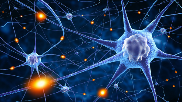
Collaborative research between multiple departments at Rutgers University and Stanford University, has resulted in the development of a 3-D micro-scaffold capable of promoting stem cells to differentiate into nerve cells. In addition, the scaffold technology supports the formation of connections between the human neurons.
Compared to transplantation of individual cells into mouse brains, the connected cells had a higher rate of survival post-injection. This new platform could pave the way for neuronal transplants to be used to treat a myriad of neurodegenerative disorders.
Neuron transplants as a viable treatment option for patients with conditions like Parkinson’s disease, have been held back by the low survival rate of the injected cells. The current study – published in the journal, Nature Communications – describes a new technique for cell culture using a 3-dimensional tissue scaffold.
“Working together, the stem cell biologists and the biomaterials experts developed a system capable of shuttling neural cells through the demanding journey of transplantation and engraftment into host brain tissue,” said Dr. Rosemarie Hunziker, director of the National Institute of Biomedical Imaging and Bioengineering (NIBIB) Program in Tissue Engineering and Regenerative Medicine. “This exciting work was made possible by the close collaboration of experts in a wide range of disciplines.”
In order to determine the optimal design for the tissue scaffold, the researchers made prototypes using a number of different variations of polymer fibers, each of which varied in terms of density and thickness. They found that the stem cells optimally adhered to a web-type structure composed of relatively thick polymer fibers.
The researchers used human induced pluripotent stem cells (iPSCs) for the study, which are often generated from adult skin cells. The addition of the protein NeuroD1, was responsible for inducing the stem cells to differentiate into neural cells.
According to the researchers, one of the most critical aspects of the scaffold was the spacing between adjacent polymer fibers. “If the scaffolds were too dense, the stem cell-derived neurons were unable to integrate into the scaffold, whereas if they are too sparse then the network organization tends to be poor,” said Dr. Prabhas Moghe, a professor of biomedical engineering and chemical engineering at Rutgers University, and a senior author on the publication.
“The optimal pore size was one that was large enough for the cells to populate the scaffold but small enough that the differentiating neurons sensed the presence of their neighbors and produced outgrowths resulting in cell-to-cell contact,” said Moghe. “This contact enhances cell survival and development into functional neurons able to transmit an electrical signal across the developing neural network.”
The micro-scaffolds developed by the team were small enough to fit inside a standard hypodermic needle, which was used to inject them into the mouse brains. The researchers also injected dissociated neurons into other mice, and compared cell survival by analyzing thin slices of the brain tissue.
The researchers found that cell survival for scaffold-grown neurons was almost 40 percent higher compared to cells grown in solution. Critically, the scaffolds improved electrical activity, neuronal outgrowth and maturation of synapses between neurons, suggesting that the transplanted cells were integrated well with the endogenous brain cells.
The team has plans to apply the technology as a future treatment for Parkinson’s disease. To do this, the researchers will need to refine the technique to induce stem cells to differentiate into dopamine-producing neurons – the neuronal cell type lacking in patients with Parkinson’s.


Join or login to leave a comment
JOIN LOGIN