Video of heart drugs on human cardiac muscle cells housed in an inch-long silicone device. This video shows muscle cells before and after exposure to isoproterenol, a drug used to treat certain heart ailments, including bradycardia (slow heart rate). It’s clear that the cells beat faster after 30 minutes of exposure to isoproterenol.
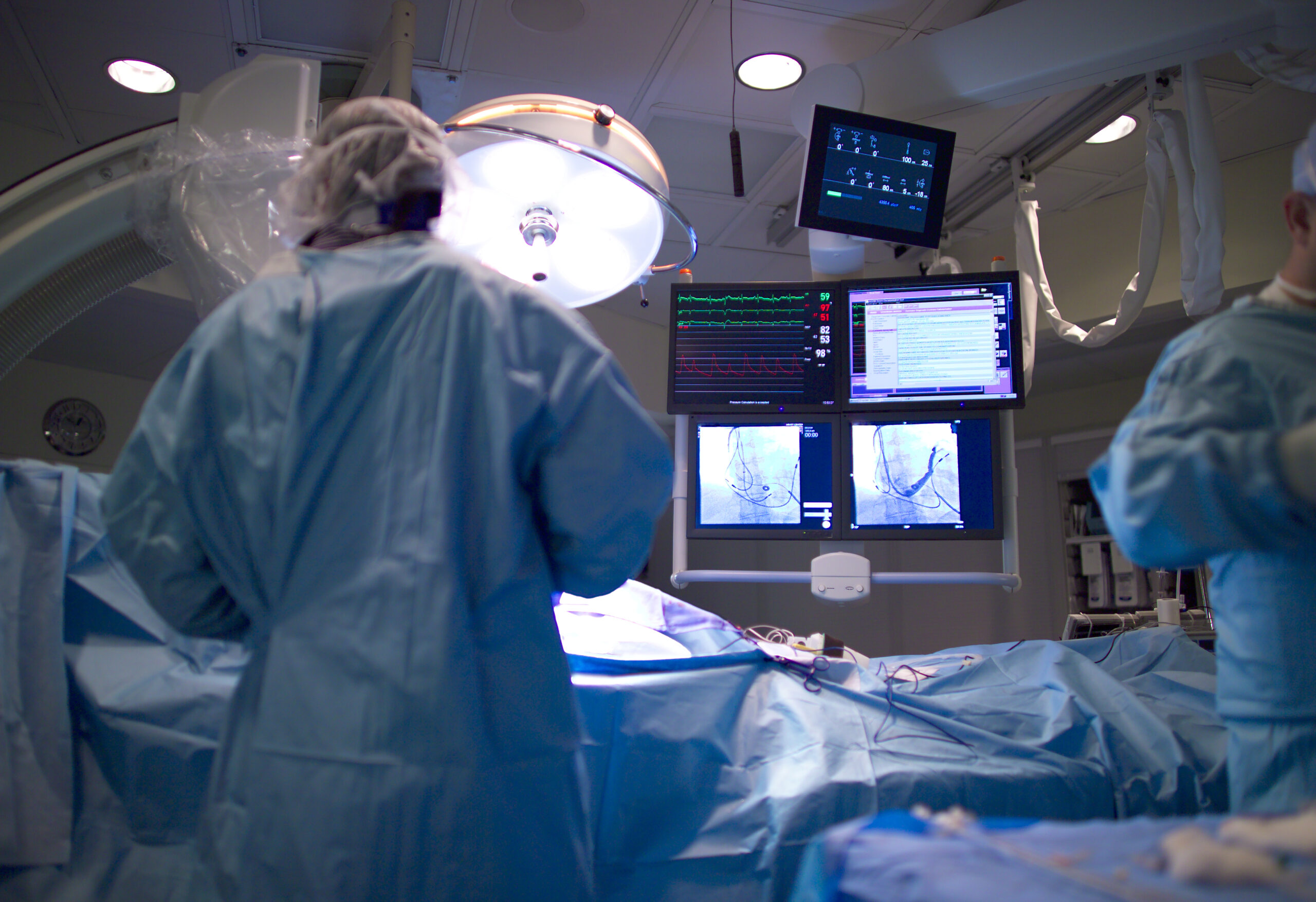
The painstakingly long drug development process (~10-15 years) that tends to cost billions of dollars is mainly attributed to the preclinical and clinical phases. It is a high risk investment considering approximately one third of pharmaceutical safety testing is withdrawn due to cardiotoxicity.
Over-simplified cell and animal models do not faithfully recapitulate the complex biological human response to drug treatment, and also have strong ethical and economic considerations. Thus, more accurate models that mimic human microenvironments are necessary to predict cardiotoxic responses, before putting human lives directly at risk and wasting large economic investments and valuable resources.
Epigenomics is a genome wide approach that identifies specific DNA sequences where these processes are targeted. Functional methylomes have been mapped using next generation capture sequencing technologies to distinguish variability in methylation patterns between cell types and disease.
Heart-on-a-Chip: Human Cardiac Microphysiological System
Dr. Kevin Healy and his research team from the University of California, Berkeley, have microfabricated a 3D tissue construct that recapitulates a minimal organoid structure composed of pulsating human myocardium – essentially, a beating heart-on-a-chip. This cardiovascular microphysiological system has shown to effectively predict the cardiotoxicity of various heart drugs, consistent with pharmacological data on tissue scale references compared to cellular scale studies.
This system utilizes the unprecedented potential of human induced pluripotent stem cells (hiPSCs) that have the ability to differentiate into any cell type. hiPSCs contain a human genetic background and can be derived from patients or genome-edited for disease studies.
In this case, differentiating cardiomyocytes confined to biomimetic dimensions have been intelligently designed to facilitate their own self-organization and structural alignment. This allows for the formation of multiple cell layers with interacting extracellular matrix components, and the eventual generation of a uniaxial, robust and homogenous beat rate. The beat rate is used as a metric for measuring drug response in a noninvasive, sensitive, and reproducible manner.
This system employs a microfluidic component to emulate the microcirculation that naturally occurs between organ and vasculature. The system is composed of a large central cell chamber with two adjacent media channels. They are connected by microchannel arrays that mimic the endothelial barrier, allowing passive ‘tissue-like’ diffusion of nutrients and drug compounds as well as protection against sheer force.
This dynamics in-vivo-like environment can more efficiently assess relevant pharmacokinetics and pharmacodynamics of drug response that are likely to occur in a human body. This novel technology has potential to revolutionize drug discovery and development, reducing both cost and duration of bringing new drug candidates to market.
We should anticipate widespread adoption of microphysiological systems of more interactive model organs, paving the way for the possibility of eventually developing a ‘human-on-a-chip’ system for drug screening and disease modelling.
Click here to read the full study in Nature Scientific Reports


Join or login to leave a comment
JOIN LOGIN