The preclinical phase of drug discovery commonly includes going through numerous histopathological samples. This contributes to the drug development process being time-consuming and labour-intensive for pharmaceutical and contract research organizations. Difficulties also stem from having to detect very subtle changes with high precision and accuracy. Manual quantification of small changes, or specific cell counting, is not only cumbersome but also, often involves high costs.
The digitization of glass slides has paved the way for even more advanced technology like artificial intelligence (AI), to further advance image analysis in a variety of medical fields. AI-based methods have the potential to standardize slide review by reducing bias while increasing the speed and accuracy of analysis.
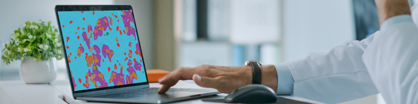
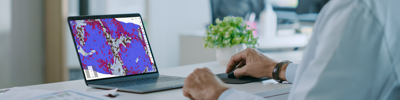
In this webinar, the featured speaker discusses utilizing a cloud-based software from Aiforia Technologies, for automating image analysis tasks with AI to enhance the CRO’s work by providing higher quality data and therefore confidence in this data to their clients across pharmaceutical companies. Through the software, the speaker created several AI models for assessing different markers from the central nervous system (CNS) tissue.
Join this webinar to hear case studies with large pharmaceutical and biotechnology clients, discover ways to harness artificial intelligence for higher quality data in preclinical trials and translational research and discuss how deep learning augments workflows, providing quantifiable benefits from CRO to client.
Speaker

Tate York, Director of Digital Image and Analysis, NSA Labs
Tate York received his master’s degree in pharmacology and toxicology from Michigan State University and has been working at NSA Labs for the past ten years. He is currently the Director of Digital Imaging and Analysis for NSA Labs.
Who Should Attend?
Professionals in contract research organizations or pharmaceutical companies including:
- Scientists, Pathologists, Principal Scientists, Toxicologists, Senior Scientist, Senior Toxicologist
- Directors of R&D or digital imaging, Lab Heads, Research and Development Coordinator, Head of Development, Head of Business Development, VP Research and Development
What You Will Learn
Attendees will learn:
- How deep learning artificial intelligence (AI) can be used in histology or pathology image analysis
- How AI augments preclinical investigation workflows
- Case studies of CROs, pharma and biotech and the benefits they experienced from using AI software
- How to create AI models without the need for coding for any image analysis task in histology or pathology
Xtalks Partner

Aiforia
Aiforia equips pathologists and scientists in preclinical and clinical labs with powerful deep learning artificial intelligence software for translating images into discoveries, decisions, and diagnoses. The cloud based Aiforia products and services aim to escalate the efficiency and precision of medical image analysis beyond current capabilities, across a variety of fields from oncology to neuroscience and more. To learn more, please visit www.aiforia.com
Related Webinars
You Must Login To Register for this Free Webinar
Already have an account? LOGIN HERE. If you don’t have an account you need to create a free account.
Create Account