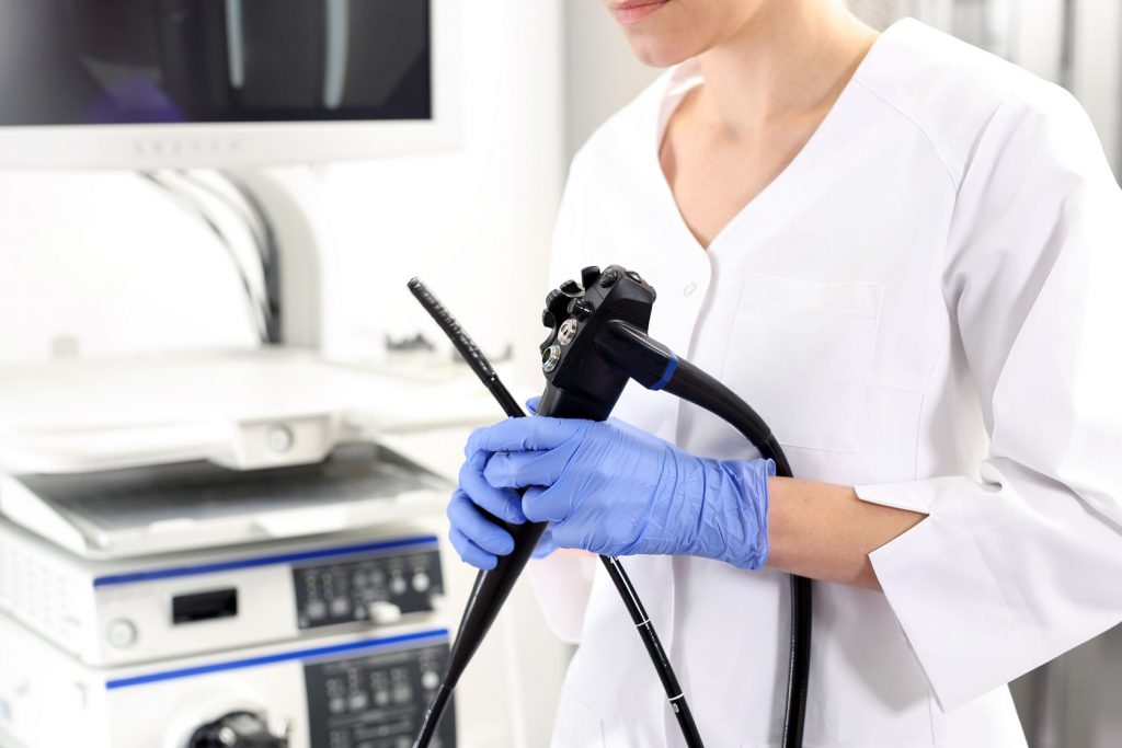
A team of researchers from Showa University in Yokohama, Japan have developed an endoscope which uses artificial intelligence (AI) to identify cancerous lesions during routine colonoscopy. The diagnostic system could improve early diagnoses of colorectal cancer.
A special type of endoscope – known as an endocytoscope – prototyped by medical device maker Olympus, was used to view colorectal poylps at 500x magnification. Using machine learning, the AI was trained to compare around 300 features of the image of the polyp to a database of more than 30,000 images.
After performing this image analysis, the AI could make predictions on whether the polyp is likely to be malignant or benign. The Japanese researchers recently tested this AI-guided imaging system in a prospective study, the results of which were presented at the 25th United European Gastroenterology (UEG) Week in Barcelona, Spain.
“The most remarkable breakthrough with this system is that artificial intelligence enables real-time optical biopsy of colorectal polyps during colonoscopy, regardless of the endoscopists’ skill,” said Dr. Yuichi Mori of Showa University. “This allows the complete resection of adenomatous polyps and prevents unnecessary polypectomy of non-neoplastic polyps.”
In all, the study involved 250 participants who were found to have colorectal polyps after undergoing imaging using endocytoscopy. The ability of the AI system to predict the pathology of these polyps was compared to standard methods for identifying colorectal cancer.
They found that the real-time AI-assisted imaging device had a sensitivity of 94 percent and a specificity of 79 percent. In addition, the diagnostic tool showed 86 percent accuracy at identifying neoplastic changes in the cells.
“We believe these results are acceptable for clinical application and our immediate goal is to obtain regulatory approval for the diagnostic system,” said Mori.
The next steps in the development of this AI-guided imaging system is to conduct a larger, multicenter study to further investigate the feasibility of using the diagnostic to detect colorectal cancer. If results continue to be promising, this tool could improve the time it takes to identify a malignant lesion, helping clinicians to remove the masses before they develop further.
“Precise on-site identification of adenomas during colonoscopy contributes to the complete resection of neoplastic lesions,” said Mori. “This is thought to decrease the risk of colorectal cancer and, ultimately, cancer-related death.”


Join or login to leave a comment
JOIN LOGIN