According to the National Institutes of Health (NIH), between two and three out of every 1,000 children in the US are born with some level of hearing loss, which can, in some cases, be corrected surgically or with the use of external hearing aids. However, commercially-available prostheses aren’t always a perfect fit, resulting in high failure rates.
To address this issue, researchers at the University of Maryland have developed custom-designed middle ear implants using CT scans and 3-D printing. They presented their research at the annual meeting of the Radiological Society of North America (RSNA).
Sound vibrations are through the ear drum to the cochlea by way of the ossicles, three small bones in the middle of the ear. These tiny bones can become damaged in a number for ways – such as infection or trauma to the ear – which results in ossicular conductive hearing loss.
Currently, doctors use ceramic cups and stainless steel struts to surgically reconstruct the middle ear damage, however they are not always successful. Customization of the prosthesis occurs in the operating room and is often limited.
“The ossicles are very small structures, and one reason the surgery has a high failure rate is thought to be due to incorrect sizing of the prostheses,” said study author Dr. Jeffrey D. Hirsch, assistant professor of radiology at the University of Maryland School of Medicine (UMSOM) in Baltimore. “If you could custom-design a prosthesis with a more exact fit, then the procedure should have a higher rate of success.”
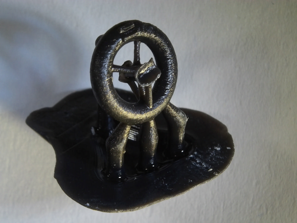
In order to create the customized, 3-D printed implants, Hirsch and his colleagues used CT imaging to model the middle linking bone in the ossicular chain removed from three human cadavers. Using the CT images, they used a 3-D printer to created custom prostheses out of a special resin designed to harden when exposed to UV light.
The 3-D printed bones were then given to four surgeons who were asked to match the prosthesis to the right recipient. All of the surgeons were able to identify which bones were custom-printed for which individuals, emphasizing the ability of this technology to overcome the challenges of this type of procedure.
“This study highlights the core strength of 3-D printing — the ability to very accurately reproduce anatomic relationships in space to a sub-millimeter level,” said Dr. Hirsch. “With these models, it’s almost a snap fit.”
Hirsch and his team are now looking to build prostheses out of biocompatible materials so that they can be tested on living patients. They are also investigating the possibility of printing a scaffold seeded with stem cells which, when implanted, could help a patient regrow the damaged tissue.
“Instead of making the middle ear prosthesis solid, you could perforate it to be a lattice that allows stem cells to grow onto it,” Dr. Hirsch said. “The stem cells would mature into bone and become a permanent fix for patients with hearing loss.”


Join or login to leave a comment
JOIN LOGIN