The use of advanced image analysis techniques with IHC assays is enabling robust quantitative analysis of biomarker expression and localization in tissue samples. In this webinar, three unique case studies will be presented that demonstrate how IHC-based assays have been used to advance basic research and preclinical studies in diabetes, immuno-oncology, and oncology:
Case Study 1: Development of an Encapsulated Cell Replacement Therapy for Diabetes
- Anindita Bhoumik of ViaCyte, Inc. will review the development of a stem cell-based islet replacement therapy for treatment of patients with diabetes. She will address how advanced image analysis enabled product characterization with single and dual IHC staining.
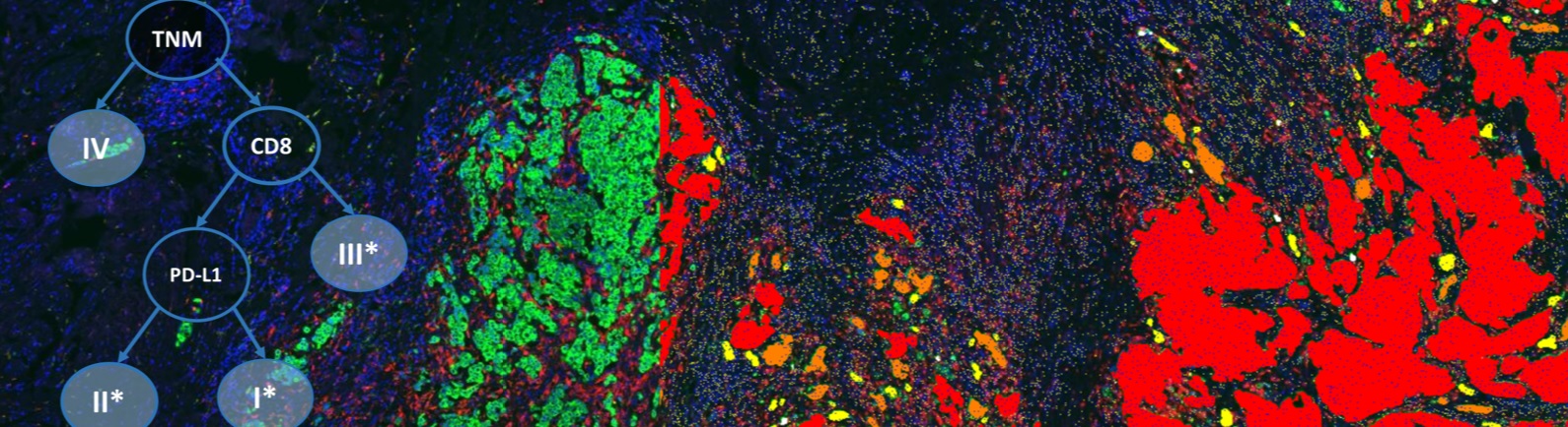
Case Study 2: An Image Analysis Algorithm Based on the Hue Saturation Density Transformation, an Important Tool for Melanoma Immunotherapy Research
- Dr. Jeff Dzubay of Definiens will review how to improve melanoma research. He will demonstrate how to successfully separate red stained CD3 and CD8 positive immune cells from a background of brown melanocytes in TMA core samples from melanoma patients.
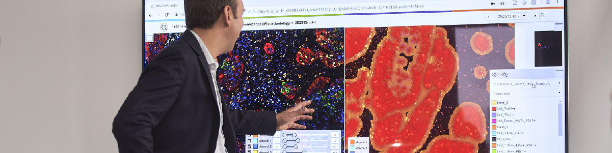
Case Study 3: Utilizing Definiens Tissue Studio® to Support Preclinical Assay Development for IHC-based Biomarkers
- Douglas Bowman of Takeda Pharmaceuticals will highlight the image analysis workflow deployed at Takeda for creating and analyzing IHC-based biomarkers. Using the PD assay workflow for context, he will highlight preclinical IHC image analysis and provide tips and tricks they have learned to get the most out of tissue image analysis.
Speakers

Anindita Bhoumik, M.S., Scientist, R&D, ViaCyte, Inc.
Ms. Bhoumik is responsible for various aspects of ViaCyte’s research and development program including pre-clinical studies, assay development and stem cell differentiation. She leads the company’s cell-product scale-up optimization and technology, transfer of projects internally and to corporate partners. Prior to joining ViaCyte, she was at Mount Sinai School of Medicine, NY and Sanford-Burnham Medical Research Institute in La Jolla, CA where she has made major contributions in the field of cancer, studying signaling networks that are often dysregulated in cancer, in an effort to understand why they are dysregulated, and how it may be possible to correct them. She is a co-author of high profile publications in peer-reviewed scientific journals and of a book chapter which describes the progression to malignancy in melanomas. She is a co-inventor on several patents related to Melanoma. Anindita earned her M.S. in Organic Chemistry from the Banaras Hindu University, India.

Jeff Dzubay, Ph.D., Field Application Scientist, Definiens
Jeff Dzubay earned his BS in Physics from the University of Washington in 1993, and his PhD in Neuroscience form the Oregon Health Sciences University in 1999. He is a Field Application Scientist with Definiens, and enjoys sharing the technology with potential customers, training new customers, and working on custom services projects. He has over twenty years of experience with microscopy and digital imaging and analysis. He has held lead scientist and program management roles developing cell-based assays for the drug discovery market, primarily using fluorescent ion-indicator dyes and related technologies.

Douglas Bowman, Associate Scientific Fellow, Takeda Pharmaceuticals International Co.
Douglas Bowman, Associate Scientific Fellow, Takeda Pharmaceuticals International Co. Douglas Bowman is currently an Associate Scientific Fellow at Takeda Pharmaceuticals where he is responsible for cell-based and tissue-based imaging technologies and assays to support multiple oncology projects. Douglas manages a group that supports both in vitro and in vivo imaging and biology efforts for assays utilizing technologies such as High Content Screening, live-cell imaging, and digital pathology whole slide scanners. He has led the deployment and development of technologies and automated tools to enable high throughput IHC-based pharmacodynamic biomarker assays to support preclinical studies. He has been an invited speaker at a number of industry conferences and vendor user group meetings.
Who Should Attend?
This webinar will be ideal for scientists in Pharmaceutical, Biotech and Academic Medical Centers who focus their research efforts in discovery and preclinical studies in the areas of oncology, immunotherapy, and metabolism.
Xtalks Partner

Definiens
Definiens is the leading provider of image analysis and data mining solutions for tissue diagnostics and clinical digital pathology. Definiens technology provides detailed cell-by-cell readouts from target structures on tissue slides and allows the correlation of this information with data derived from other sources, generating new knowledge and supporting better decisions in research, diagnostics and therapy. Definiens’ Tissue Phenomics approach was awarded the 2013 Frost and Sullivan Company of the Year Award for Global Tissue Diagnostics and Pathology Imaging. For more information, please visit: www.definiens.com.
Media Partner
Related Webinars
You Must Login To Register for this Free Webinar
Already have an account? LOGIN HERE. If you don’t have an account you need to create a free account.
Create Account