Patient biopsy samples received from Adaptimmune’s clinical trials are considered very precious, therefore careful consideration is taken to decide which histological staining and assays are used on these tissue sections. These analyses serve to achieve our team and research department’s primary endpoints as well as help determine which assays can provide the clearest understanding of the whole tumour microenvironment (TME).
Multiplex immunofluorescence (mIF) assays, like Ultivue’s 8-plex panels, offer the advantage of preserving the architectural features of the tumour microenvironment (TME) by utilising a single tissue section for multi-target profiling. This can reveal the spatial relationships between tumour cells, Adaptimmune engineered T-cells, endogenous T-cells, other immune cells, and stromal components that are present.
In this webinar, we will introduce how we use these Ultivue panels alongside conventional H&E, IHC, and our other in-house assays. In addition, we will show examples of pre- and post-treatment patient biopsies for multiple indications to demonstrate how we utilize image analysis software to quantify cell densities of subsets of infiltrating immune cells. We will also describe how we use the spatial analysis tools to study cellular spatial patterns within the tumour tissue, and how these are used to understand the effectiveness and mechanism of our cell-based therapies.
Register for this webinar today to learn how multiplex immunofluorescence panels help determine the effectiveness of cell therapies for solid tumors.
Speakers

Dr. Martin Isabelle, Associate Director, Tumour Profiling, Translational Sciences Group, Adaptimmune
Dr. Martin Isabelle received his BSc Hons degree (1999) in Biochemistry at the University of Lancaster, Lancaster, UK, and has received his PhD degree (2008) at Cranfield Postgraduate Medical School (CPMS), Cranfield University, Cranfield, UK.
He subsequently worked in several postdoctoral fellow/Research associate fellow positions for eight years in US, Canada and UK, working in medical diagnostics/imaging, bio-spectroscopy, radiobiology, theradiagnostics and immuno-oncology.
Read more...
Since November 2021, Martin has led the tumour profiling team within Translational Sciences at Adaptimmune. His team is responsible for analysing clinical tumour biopsies from Adaptimmune’s clinical programmes; detecting and quantifying the presence of Adaptimmune’s TCR T-cells; and understanding the tumour microenvironment and its contribution to the immunotherapy treatment efficacy and supporting new TME target discoveries. His team utilises multiple multiplex staining and imaging technologies, principally spatial immunophenotyping, alongside bulk and spatial genomics and transcriptomics profiling.
Read Less...
![]()

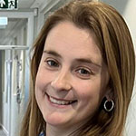
Dr. Robyn Broad, Principal Scientist, Tumour Profiling, Translational Sciences Group, Adaptimmune
Following completion of her undergraduate degree, Dr. Robyn Broad received her MSc in Cancer Research and Molecular Biomedicine from the University of Manchester and completed her PhD from the School of Medicine at the University of Leeds in 2019 where she investigated the role of cancer-associated fibroblasts on chemoresistance in triple-negative breast cancer.
Since 2018, Robyn has worked at Adaptimmune, firstly within the Target Validation team for identifying new targets to add to the Adaptimmune pipeline. Having cross-trained within the Translational Team during her time in Target Validation, Robyn moved over to the Translational Sciences team in January 2020 and joined the Tumour Profiling team.
As part of the Tumour Profiling team, Robyn is responsible for the analysis of tumor biopsies from Adaptimmunes’ clinical trials, identifying the presence of TCR T-cells and understanding the tumour microenvironment within tumour biopsies. This is done through the use of multiplex staining and imaging technologies with spatial immunophenotyping alongside genomics and transcriptomics profiling.

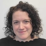
Devan Fleury, Senior Image Analysis Scientist, Ultivue
Devan Fleury received her Bachelors in Animal Science and Masters in Applied Genomics from the University of Connecticut. She has worked for over 13 years in spatial image analysis in both academia and industry. Devan began her career as a bench scientist at Boehringer Ingelheim working in the Immunology and Respiratory Disease Research molecular histopathology lab. She developed IHC, IF and ISH assays as well as implementing many commercial methods of spatial transcriptomics and proteomics. Besides the benchwork, Devan developed a fascination with image analysis and developing new methods for visualizing histology data at BI which led to her moving to Ultivue in 2021. Devan has worked at Ultivue for 2 years as a Senior Image Analysis Scientist.
Who Should Attend?
This webinar will appeal to:
- Pathologists, research pathologist professionals, image analysts and immunotherapy medical professionals who are using or want to use multiplex tissue-based assays and image analysis software
- Computational biologists who want to understand some of the techniques used to phenotype and quantify cell types in tissues and utilise spatial analytic methods
What You Will Learn
Attendees will learn about:
- How mIF is being used to evaluate the effectiveness of immunotherapy clinical trials
- Utilising advanced spatial metrics analysis tools to study cellular spatial patterns within the tumour tissue, pre and post-treatment, to understand the effectiveness and mechanism of cell-based therapies
- Using these mIF panels alongside conventional histology, multiplex IHC and other spatial proteomic and transcriptomic platforms
Xtalks Partner
You Must Login To Register for this Free Webinar
Already have an account? LOGIN HERE. If you don’t have an account you need to create a free account.
Create Account


