A new 3-D, colour medical scanner called the MARS spectral x-ray scanner has been designed to improve diagnosis of a range of diseases by providing a more detailed image of the body. Developed by father and son scientists Professors Phil and Anthony Butler, the MARS x-ray scanner will be put to use on patients who visit the Christchurch orthopedic and rheumatology center in New Zealand in the following months.
“X-ray spectral information allows health professionals to measure the different components of body parts such as fat, water, calcium, and disease markers. Traditional black-and-white x-rays only allow measurement of the density and shape of an object,” said Anthony Butler, radiologist and professor at both the Universities of Otago and Canterbury.
Using the same technology employed by the European Organization for Nuclear Research (CERN) to study the Higgs boson (also referred to as the ‘God particle’), the Butlers created a medical scanner which outperforms current devices in use today. Their MARS CT scanner produces images in full colour by measuring the x-ray spectrum which allows physicians to view multiple body tissues and components, including lipids, water, calcium and even disease biomarkers.
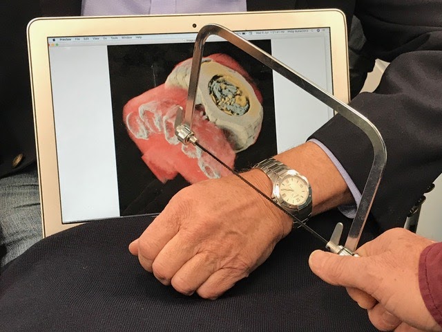
While pared-down versions of the scanner are currently in use to study tissue samples, Professor Phil Butler became the first human to have parts of his body – namely, his wrist and his ankle – imaged by the machine. Patients at the Christchurch site will become part of a clinical trial of the technology to compare it to what is currently being used in hospitals.
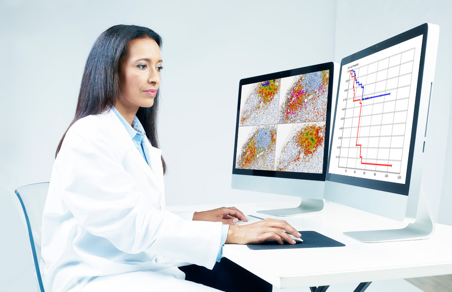
“So far researchers have been using a small version of the MARS scanner to study cancer, bone and joint health, and vascular diseases that cause heart attacks and strokes,” said Butler. “In all of these studies, promising early results suggest that when spectral imaging is routinely used in clinics it will enable more accurate diagnosis and personalisation of treatment.”
The technology is being further developed and commercialized by MARS Bioimaging, a company run by the Butlers. The MARS x-ray scanner was 10 years in the making.
“As a new imaging device, a new microscope if you like, biomedical researchers can non-invasively see different kinds of detail inside patients,” said Butler.


Join or login to leave a comment
JOIN LOGIN